


MRI of the Pelvis
COMPARISON: MRI of Pelvis 11/06/2017
TECHNIQUE: Multiplanar images of the pelvis were obtained at 1.5 Tesla prior to and following administration of 8 mL of Gadavist intravenous contrast.
FINDINGS:
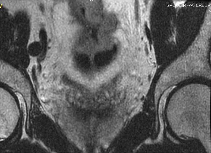
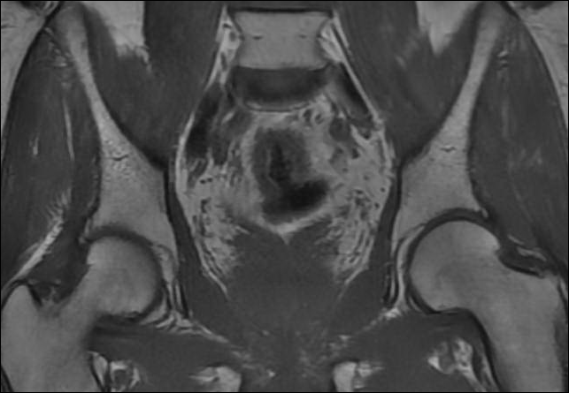
GI TRACT: There is a concentric, heterogeneous lobular mass along the wall of the rectosigmoid junction extending over length of 5.9 cm and measuring up to 1.6 cm in thickness. The distal extent of this lesion is approximately 13.1 cm proximal to the anal verge. There is enhancing extra serosal tissue along the left and right lateral walls of the thickened segment of bowel consistent with extra serosal spread of tumor. Compared to the previous MRI, the left lateral extra-serosal soft tissue mass is decreased in size. The maximal wall thickness on the prior study measured approximately 3 cm in thickness.
URINARY BLADDER: There is diffuse bladder wall hypertrophy. No bladder mass is seen.
PROSTATE: The prostate is normal in size and signal.
SEMINAL VESICLES: The seminal vesicles are symmetric bilaterally and appear unremarkable.
LYMPHATICS: There is an 8 mm right external iliac chain lymph node which is unchanged. No pelvic lymphadenopathy is otherwise seen.
BONES: There is no fracture or bone lesion.
IMPRESSION: Interval decreased thickness of the rectosigmoid mass in comparison to 11/6/2017. There is extra-serosal extension of tumor into the perirectal fat. Stable right external iliac 8 mm lymph node. No pelvic adenopathy or additional evidence of metastatic disease.
For more information on MRI imaging services at Greater Waterbury Imaging Center, visit our clinical section of the website.