MRI Pelvis Prostate Exam with and without IV contrast
HISTORY: HIGH GRADE PROSTATIC INTRAEPITHELIAL NEOPLASIA, ELEVATED PROSTATE SPECIFIC ANTIGEN
COMPARISON: CT 6/26/2013
TECHNIQUE: Multiplanar images of the pelvis were obtained at 1.5 Tesla prior to and following administration of 8 ml. of Gadavist intravenous contrast.
FINDINGS:
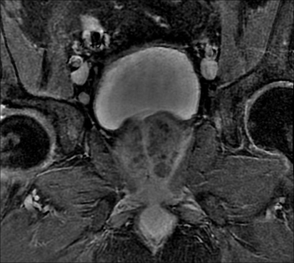
Urinary Bladder: The urinary bladder appears normal, without stones or wall thickening.
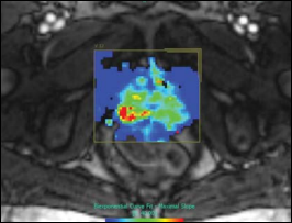
Prostate: The prostate gland is enlarged, with marked benign prostatic hypertrophy centrally. The prostate measures 5.5 cm transversely x 4.4 cm AP x 5.1 cm craniocaudally, for an overall volume of 64 cc. At the time of the gland laterally on the left, within the peripheral zone, there is a poorly marginated region of decreased signal, with focal diffusion restriction, suspicious for tumor, measuring 2 cm in diameter. No other areas of definite soft tissue abnormality are identified. There is some distortion of the left lateral capsular margin of the prostate gland, raising a possibility of capsular invasion. The neurovascular bundle has a normal appearance bilaterally.
Seminal Vesicles: The seminal vesicles are symmetric bilaterally and appear unremarkable.
Lymphatics: No pelvic lymphadenopathy is present.
Bones: There is no fracture or bone lesion.
IMPRESSION: 2 cm region of decreased signal, with some mass effect and diffusion restriction involving the left lateral aspect of the prostate gland at the base, suspicious for tumor. Distortion of the lateral capsular margin of the prostate gland could be related to capsular invasion. No other evidence of extracapsular spread of tumor.
PIRADS 4: High (clinically significant cancer is likely to be present)