MRI Thoracic Spine Case Study
HISTORY: Trauma
COMPARISON: CT 5/28/2018
TECHNIQUE: Axial and sagittal images of the thoracic spine were obtained at 1.5 Tesla
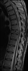
FINDINGS: As noted on the recent CT scan of the chest, there is a compression fracture of the T8 vertebral body, with loss of vertebral height by about 50%. There is retropulsion of bone at the level of the compression fracture, narrowing the AP diameter of the thoracic spinal canal by 40%. There is slight posterior displacement of the thoracic spinal cord, without cord compression. At the level of the T8 compression fracture, there is abnormal bone marrow signal, consistent with an underlying hemangioma. There is no abnormal signal within the visible spinal cord. There are no intradural abnormalities. There is no paravertebral mass or fluid collection. No thoracic disc herniation. There are small bilateral pleural effusions, with atelectasis at the lung bases posteriorly. Cerebral hepatic lesions are again noted, as seen on the recent CT scan.
IMPRESSION: Recent T8 vertebral compression fracture, with posterior protrusion of bone causing a moderate degree of central spinal stenosis, slightly displacing the spinal cord posteriorly but without cord compression.