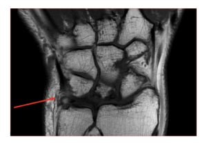
Our case study of the month is an MRI scan of the left wrist. The MRI Wrist Case Study procedure included multiplanar images of the left wrist which were obtained on our 1.5 Tesla MRI machine.
MRI Wrist Case Study Exam Findings
History: Left Wrist Osteoarthritis
Comparison: None
Technique: Multiplanar images of the left wrist were obtained at 1.5 Tesla without IV contrast.
Findings
Bones: There is old degenerative fragmentation of the lunate, with multiple cysts throughout the lunate and several small bone fragments along its lateral and dorsal margins. There are multiple degenerative cysts within the triquetrum as well There are osteophytes and degenerative subcortical cysts in the distal radius, adjacent to the articulation with the lunate. Not evidence of acute fracture.
Muscles and Tendons: There is normal signal in the visualized muscles and tendons. The flexor retinaculum is intact.
Ligaments: No evidence of ligament rupture is seen. The triangular fibrocartilage complex is intact.
Soft Tissues: There is normal signal in the visualized soft tissues. No mass or abnormal fluid collection is seen.


