

Visionaire Cutting Blocks Image Quality Checklist
Magnetic Resonance Imaging for VISIONAIRE™ Patient Matched Instrumentation by Smith & Nephew, Inc.
Greater Waterbury Imaging Center is one of a handful of imaging centers in Connecticut certified to provide specific MR images of the knee anatomy for patients who need knee joint replacement and are using the VISIONAIRE™ product. The precision of the images for the Visionaire Cutting Blocks is critical to the success of the patient’s surgery and we are proud to be able to provide these scans.
Imaging and Image Quality Checklist
As the precision of the images is critical to the success of the patient’s surgery, GWIC adheres to the following protocol to make sure the images sent to VISIONAIRE™ meet all their requirements to produce the required product (images provided courtesy of VISIONAIRE™ Patient Matched Instrumentation by Smith & Nephew, Inc.).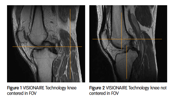
- The knee should be centered in the field of view (FOV) and if there is any coil cut-off on the images, this should be evenly distributed between superior and inferior margins of the FOV (Figures 1 and 2).
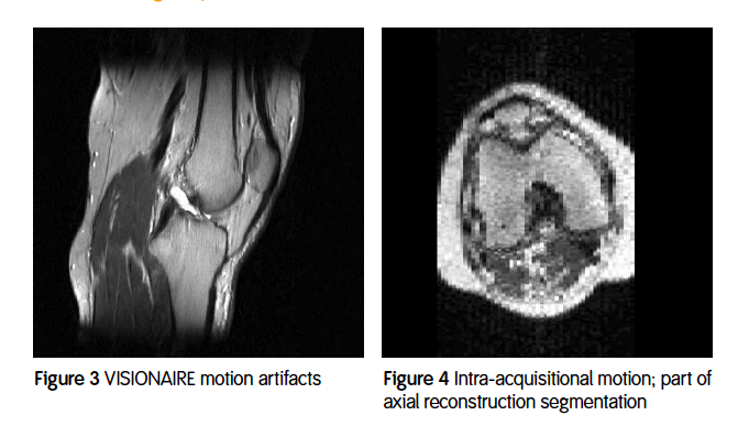
- No motion on the image set can be accepted. This includes motion that caused actual image quality degradation (Figure 3) and intra-acquisitional motion (Figure 4). Intra-acquisitional motion is motion between slices that causes the image data sets to be offset from one another. This cannot be accepted because VISIONAIRE™ is using these images to create a 3D mode
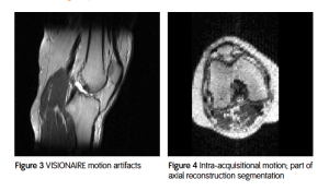 l for the Visionaire Cutting Blocks. If this type of motion is present, then the slices will not align and accurate cutting blocks cannot be made.
 - The left or right offset of the acquired image can be no more than what our scanner was approved for. This location is measured from the center of the knee or center slice.
- Our scanner has 3D distortion correction filters thus the image data that is sent must have this applied.
- The signal to noise must be good enough to visualize on all slices the cortical bone edge, the cartilage edge and the cartilage interface between the femoral and tibial cartilage.
- We must have the stated resolution on our protocols:
- 22cm FOV of 220mm FOV
- Slice thickness of 2mm
- Slice gap of 0 or interleaved
- Acquisition matrix of 512 x 256
- Reconstruction matrix must be 512 x 512
- The scan must cover the entire bony knee. Smith & Nephew usually suggests 1 or 2 slices out of bone on either side. The images must be sagittal to the patient’s knee in all three planes. Patients are more comfortable in a slightly externally rotated position in the coil. The technologist must remember to oblique the FOV in this plane also to be sagittal to the knee. Scan slices should be obliqued so that the resulting slices are perpendicular to a line drawn across the posterior femoral condyles.
For more information on the VISIONAIRE™ Cutting Blocks and the requirements for the imaging center please visit the Smith & Nephew website.