

 Breast MRI uses Magnetic Resonance Imaging (MRI) to look specifically at the breast. It is a non-invasive procedure that doctors can use to determine what the inside of the breast looks like without having to do surgery or flatten the breast (as in a mammogram).
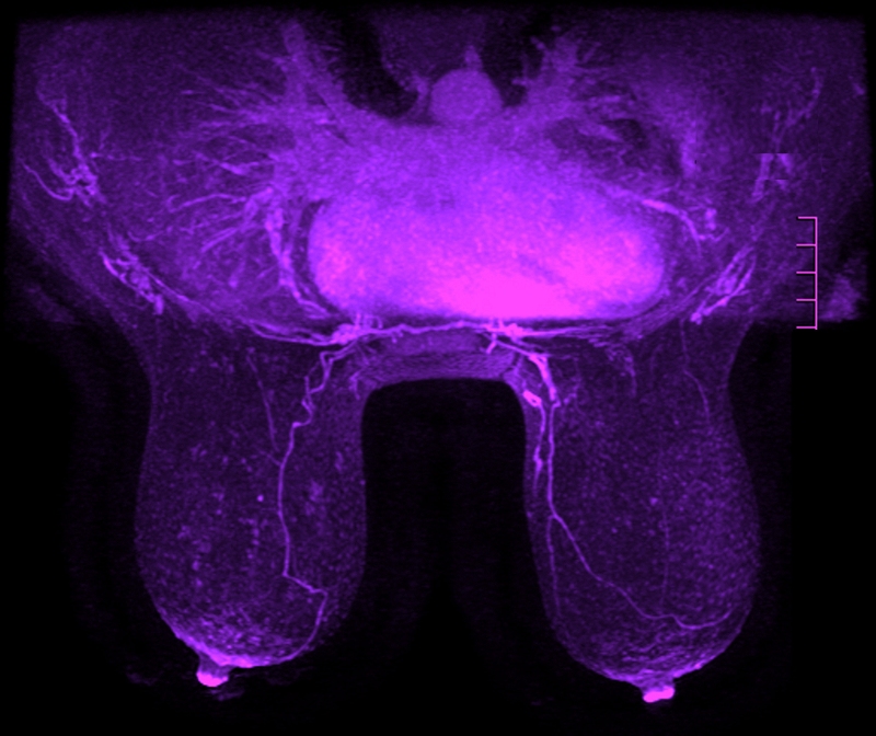
Each exam produces hundreds of images of the breast, cross-sectional in all three directions (side-to-side, top-to-bottom, front-to-back), which are then read by a Radiologist. No radioactivity is involved, and the technique is believed to have no health hazards in general. The hope is that such non-invasive studies will contribute to our progress in learning how to predict the behavior of tumors, and in selecting proper treatments.
Breast MRI is an evolving technology and should not replace standard screening and diagnostic procedures (clinical and self exams, mammogram, fine needle aspiration or biopsy). The recent interest in MRI of the breast follows reports that malignant (or cancerous) lesions get brighter following a contrast agent injection. MRI has been shown to detect small breast lesions that are sometimes missed by mammography, and MRI can successfully image the dense breast (usually found in younger women) and breast implants.
However, contrast MRI sometimes has a hard time distinguishing between carcinoma and benign breast disease. Some benign breast tissues (such as fibroadenomas) can also get bright after contrast injection, which can cause a false positive result. Research is currently going on at multiple institutions to improve breast MRI.
For MRI of the breast, the patient lies on her stomach with both breasts hanging freely into a cushioned recess containing the signal receiver (also known as the breast coil). The entire bed on which she is lying is advanced into the opening of the magnet (a tube-like machine that looks like a giant donut–open at both ends). The subject will be asked to lie still for up to 15 minutes at a time while the computer acquires the images; the total examination is made up of several scans, usually 5 to 15 minutes in length and the patient is usually in the magnet for 40-60 min.
What is the difference between Breast MRI and Mammography? From the Johns Hopkins Breast MRI Site: “Normal mammograms use x-rays to generate images of the breast tissue to search for cancer. MRI, on the other hand, uses no x-rays. The ability to identify a mass in the breast requires that the mass has a different appearance (or a different contrast) from normal tissue. With MRI, the contrast between soft tissues in the breast is 10 to 100 times greater than that obtained with x-rays.
 This is why MRI is used much more than, for example, CT (or CAT) scanning, which uses x-rays, for diagnosing tumors in the brain. As opposed to x-rays, which are known to cause damage to DNA of cells, the magnetic fields and radiowaves used with MRI are not known to have any long term biologic effect. MRI of the breast does require intravenous injection of a ‘contrast agent,’ which helps highlight breast abnormalities.
Here are more resources on when and why Breast MRI is done for breast cancer detection:
https://greaterwaterburyimagingcenter.org/magnetic-resonance-imaging/breast-mri/
https://greaterwaterburyimagingcenter.org/october-is-national-breast-cancer-awareness-month/
http://ww5.komen.org/BreastCancer/BreastMRI.html
http://www.radiologyinfo.org/en/info.cfm?pg=breastmr
http://www.breastcancer.org/symptoms/testing/types/mri
http://www.breastcancer.org/symptoms/testing/types/mri/diagnosis
https://www.nlm.nih.gov/medlineplus/ency/article/007360.htm