

Case Studies
MRI Images
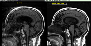 Utilizing our Propellor software you can scan patients who have difficulty holding still. In this comparison image, the MRI of the brain on the left is blurred due to patient motion. That same image, using Propellor, the software corrects the image motion and you are able to scan the patient without having to repeat scans.
Benefit: Shorter scan time, detailed imaging and patient comfort.
Lumbar Spine Case Study
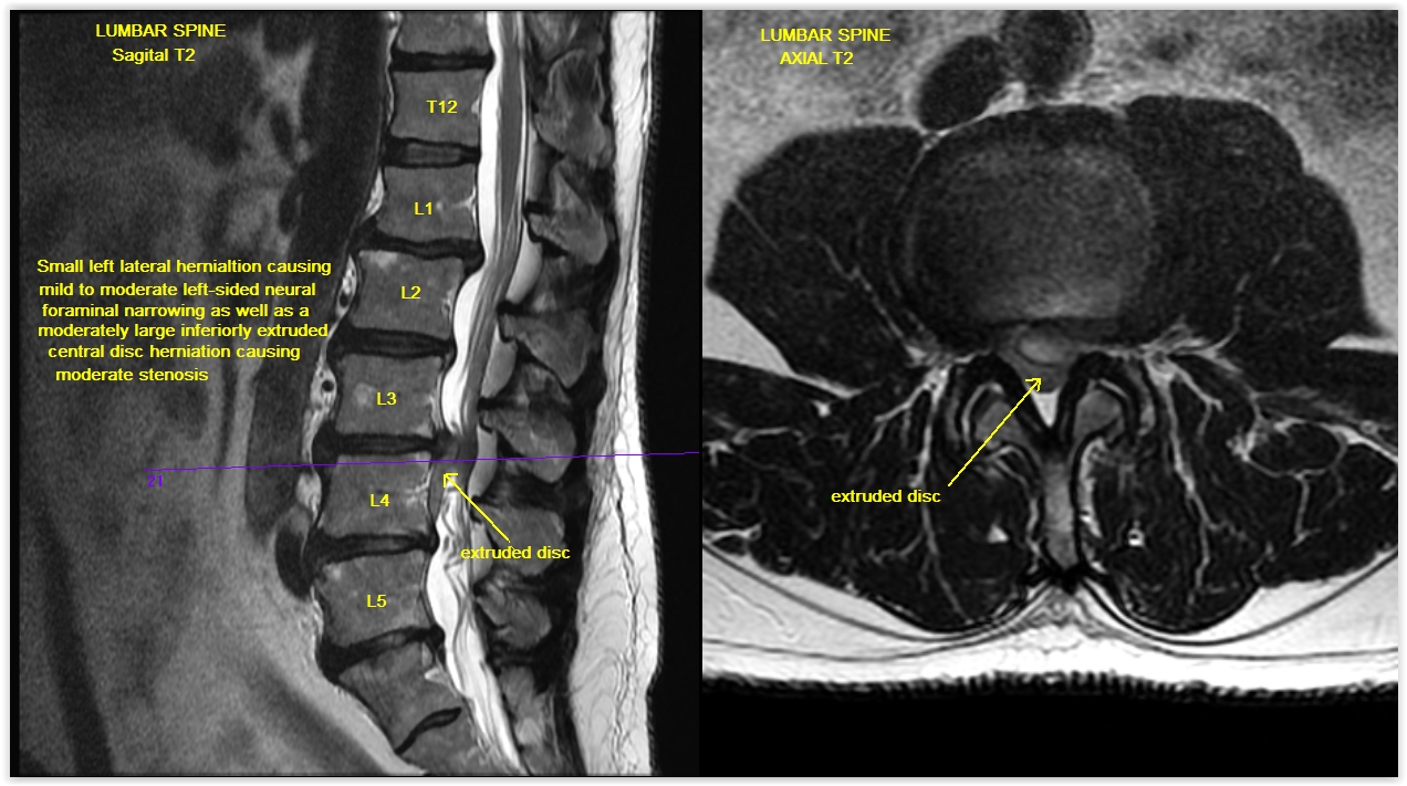 This case study is an lumbar spine Magnetic Resonance Imaging scan. The patient is a male with a history of lumbar radiculitis. This MRI Lumbar Spine Case Study procedure included axial and sagittal images of the lumbar spine. They were scanned on our 1.5 Tesla MRI machine. For all the detail on this case study click here.
MR Arthrogram Shoulder
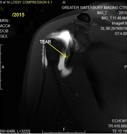 This case study is a Magnetic Resonance Arthrogram of the right shoulder or MR Arthrogram Shoulder. This 29 year old female patient presented with symptoms of chronic right shoulder pain and has a history of surgery of the right shoulder. As is seen in the image, the Magnetic Resonance exam shows a small tear of the anterior-inferior labrum. For the complete case study click here.
MR Hand Study
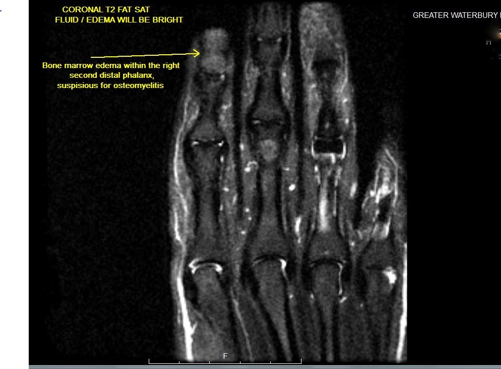 Our case study of the month is an MRI scan of the right hand. The patient presented with soft tissue swelling. The MRI Hand Case Study procedure included coronal, axial, and sagittal images of the right hand with attention to the right second digit and they were obtained on our 1.5 Tesla MRI machine. There was no comparison scan. For the complete case study click here.