

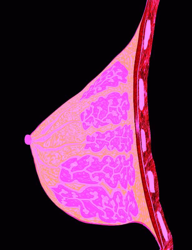
 MRI is the term which describes magnetic resonance imaging, a method used to obtain detailed images of a person’s bones, body structure, soft tissues and organs. By using a magnetic field with radio waves and computer generation, three-dimensional cross sections can be obtained to evaluate medical conditions in an individual’s body and head. MR imaging provides a valuable look inside the body that is not available with other imaging methods such as ultrasound, X-ray and computed tomography (CT) scans.
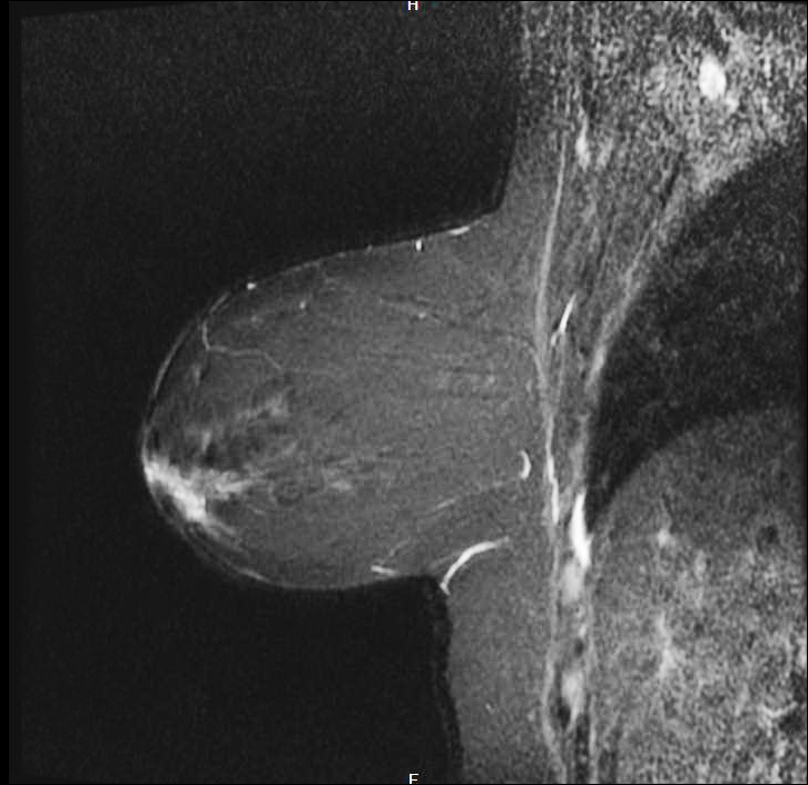
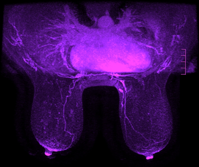
MR imaging is a valuable tool for physicians to diagnose many health conditions such as heart and vascular disease, musculoskeletal disorders, cancer, stroke and many other health conditions. MRI Breast Imaging is used to diagnose breast abnormalities, determine staging of breast cancer and is a supplement to mammography. MRI should not be considered a replacement for mammography, although may detect lesions in women with dense tissue or breast implants which can be missed during traditional mammograms.
MR imaging provides additional screening options throughout the year for women at a high risk of developing breast cancer with safe methods that do not use radiation. This makes it very safe for women of all ages.
Common Uses of MRI Breast Imaging
MRI is used for many diagnostic and treatment purposes, including the following:
- Further diagnosis of abnormalities detected in mammography
- Identify breast cancer for women at high risk, with dense tissue or breast implants
- Determine the integrity of breast implants
- Identify location and progression of any tumors
- Evaluate progress of chemotherapy
- Distinguish between recurring tumors and scar tissue
- Assess whether cancer has spread beyond the chest wall or surgical site
- Many other diagnostic purposes not available with other methods
MRI of the breast can be taken prior to and after the use of an additional contrast material which may provide additional information to physicians which helps them in diagnosing benign or malignant tumors and examining lymph nodes.
Breast Cancer Screening Recommendations
 The American Cancer Society provides the following recommended guidelines for breast cancer screening as it is important to find breast cancer early. Early detection is critical to successful treatment, with the goal of screening to diagnose breast cancer before symptoms appear. Regular screenings will detect breast cancer while it is small and still confined to the breast area which improves a woman’s prognosis.
Women of average risk include those that:
- Have no personal history of breast cancer
- Do not have a strong family history of breast cancer
- Do not carry the gene BRCA which increases risk of breast cancer
- Had no chest radiation therapy before the age of 30
Women within the average risk category should begin mammogram screenings between the age of 40 to 44, with annual mammograms between the ages of 45 to 54.
Breast Cancer Screening Recommendations for High Risk Patients
Women considered to be high risk for breast cancer should have a breast MRI and a mammogram each year beginning by the age of 30. Women in the high risk category include women who:
- Have a personal history of breast cancer
- Had chest radiation therapy between the ages of ten and thirty
- Have a lifetime risk based on family history of 20% – 25% or more
- Carry the BRCA1 or BRCA2 gene mutation determined during genetic testing
- Have a first degree relative such as a sister, brother, child or parent with the BRCA1 or BRCA2 genes, whether they have had testing or not
- Have the Bannayan-Riley-Ruvalcaba, Cowden or Fraumeni syndrome, or a first degree relative with either of these syndromes.
Visit the American Cancer Society for more information on Breast Cancer Early Detection and Diagnosis.
What to Expect During MRI of the Breast
 During an MRI of the breast you will be lying face down with the moveable bed moved into the magnetic area of the unit. You will need to lie completely still while the MRI is obtaining images, which occurs in sequences of one to fifteen minutes. You’ll be able to hear the technologist and they’ll be able to hear you during the exam. The MR technologist will do everything possible to make you comfortable, you can usually relax in between sequences.
When a contrast material is used, an intravenous (IV) line administers the contrast prior to a series of images. Your IV will be removed upon examination of your completed images, with a full analysis and report performed by the radiologist. The radiologist is trained in analyzing MRI images and provides a full report to your health care provider.
MRI and Breast Biopsy
The radiologist may recommend further testing such as a breast biopsy of suspicious tissue. An MRI guided breast biopsy allows for further testing to determine if the tumor is cancerous. A thin needle is inserted into the tissue with MR images and sent for further testing. MR imaging is an important part of breast cancer prevention and treatment, offering physicians a way to diagnose and treat patients for early detection and an improved prognosis for survival.
Greater Waterbury Imaging Center offers a clean and modern MRI facility, providing friendly and professional MR imaging services. We encourage you to know your risk for developing breast cancer and schedule all of your preventive maintenance screening tests. Contact us for all your MR imaging needs including MR imaging of the breast.