
 HISTORY: Liver disease, unspecified
COMPARISON: None.
TECHNIQUE: Multiplanar images of the abdomen were obtained at 1.5 Tesla prior to and following the administration of IV contrast.
CONTRAST: 12 mL ofDotarem intravenous contrast.
FINDINGS:
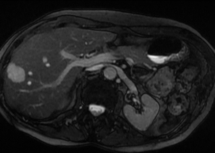
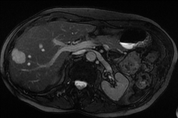
Liver: The liver is normal in size and contour. Within the posterior segment of the right lobe of the liver laterally, there is a lobular 2.4 cm soft tissue mass. After contrast administration, this lesion demonstrates a characteristic pattern of peripheral contrast enhancement, with gradually increasing enhancement centrally on the more delayed postcontrast images, typical for a hemangioma. More superiorly, in the right lobe posteriorly, there is a smaller similar 9 mm lesion. Another 6 mm enhancing nodule is present in the lateral segment of the left lobe, adjacent to the stomach.
Gallbladder: The gallbladder appears normal, without gallstones, wall thickening or
pericholecystic fluid.
CBD: The common bile duct is normal in caliber.
Pancreas: The pancreas appears normal.
Spleen: The spleen appears normal.
Adrenals: The adrenal glands are normal.
Kidneys: There is no renal mass or hydronephrosis. There is a simple appearing cystic renal lesion in the left kidney. No imaging follow-up is recommended. G9548.
Aorta: No evidence of aortic aneurysm or dissection is seen.
Lymphatics: No abdominal lymphadenopathy is present.
IMPRESSION: 2.4 cm right hepatic lesion demonstrates delayed and prolonged contrast
enhancement, typical for a hemangioma, with two other smaller subcentimeter similar lesions in the posterior segment of the right lobe and lateral segment of the left lobe.