


MR Arthrogram Case Study
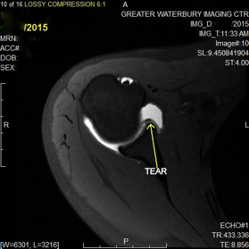
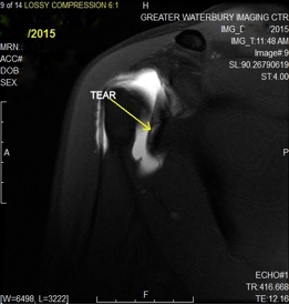
Our case study of the month is an MR Arthrogram of the right shoulder. The patient is a 29 year old female with symptoms of chronic right shoulder pain with a history of right shoulder surgery. The MRI scan shows there is a small tear of the anterior-inferior labrum.
MR Arthrogram Procedure:
- MRI of the shoulder is performed without contrast material
- The images are evaluated by the radiologist
- The patient’s physician may order an MR Arthrogram of the shoulder if a tear is suspected
- For the MR Arthrogram, contrast is injected into the shoulder joint space under fluoroscopic (x-ray) guidance
- This allows the radiologist to better see the structures in the shoulder joint
- When the contrast agent is seen in the joint space under fluoroscopic guidance, the MR scan is performed on the patient
- If there isn’t a tear, the contrast material should remain within the shoulder joint space and be absorbed into the tissue
- If the contrast leaks out of the joint space, then the radiologist can determine the size and extent of any tears or other issues
Click here for more detail on how an MR Arthrogram is done please visit our website page on MRI Shoulder Imaging.