
HISTORY: MALIGNANT NEOPLASM of RIGHT LUNG
COMPARISON: CT chest 9/9/2022
TECHNIQUE: Multiplanar images of the chest were obtained at 1.5 Tesla prior to and following administration of IV contrast. During this public health emergency, we are using enhanced sterilization processes, social distancing measures and PPE for your protection.
CONTRAST: 20 mL of Dotarem intravenous contrast
FINDINGS:
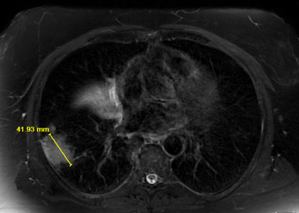
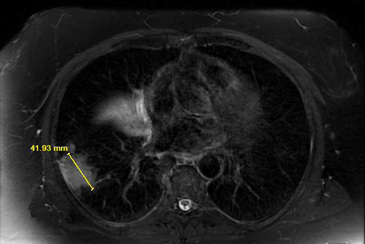
Lungs: There are multiple, scattered lung nodules consistent with metastatic disease. The largest pulmonary lesion is seen along the right posterolateral pleural surface measuring 4.2 cm in length. There also appears to be a right infrahilar mass with adjacent atelectatic lung. The infrahilar mass appears to measure up to approximately 3.6 cm in length. No significant pleural effusion.
Mediastinum: No axillary or mediastinal adenopathy. No left hilar adenopathy. The heart size is normal. The thoracic aorta is normal in caliber.
IMPRESSION:
Right lower lobe subpleural mass in the right lower lobe consistent with primary lung malignancy or additional lung nodules bilaterally in a right infrahilar mass. There is an enhancing lesion within the T9 vertebral body also most consistent with metastatic disease.