

 Reason For Exam
(MR Hips w wo Left) Pyogenic arthritis, unspecified
Report
MR LEFT HIP WITH AND WITHOUT IV CONTRAST:
HISTORY: Pyogenic arthritis, unspecified COMPARISON: 7/17/2023, 7/22/2023
TECHNIQUE: Multiplanar images of the left hip were obtained at 1.5 Tesla prior to and following the administration of 11 ml of Dotarem intravenous contrast.
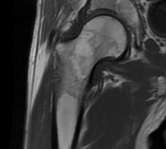
FINDINGS: As noted on the previous exam, there is significant pattern of metallic magnetic susceptibility artifact associated with internal fixation hardware transfixing a healing fracture of the proximal left femur, with a left femoral intramedullary rod. The quality of the exam is limited by patient motion artifact.
Compared to the prior exam, the pattern of abnormal bone marrow signal with edema in the left ischium has improved significantly. No new area of bone marrow edema. No evidence of osteomyelitis. There is persistent edema and fluid tracking along the left psoas muscle, but this pattern has diminished since the prior study. There is soft tissue edema within the left gluteal soft tissues, with some small loculations of fluid just deep to the left tensor fascia lata, lateral to the greater trochanter of the left femur. These fluid collections are more organized now than on the previous exam, and extend into the subcutaneous soft tissues of the left thigh laterally. There is enhancement along the periphery of these collections, raising the possibility of infection.
IMPRESSION: Left femoral internal fixation hardware artifact partially obscures the proximal left femur. No evidence of osteomyelitis.
Progressive organization of loculated fluid collections in the soft tissues lateral to the left hip, extending into the subcutaneous soft tissues of the proximal left thigh laterally, are suspicious for infected collections.
Previously noted inflammatory edema and fluid associated with the left psoas muscle are improved on this exam compared to the prior study.