
 MR PELVIS WITH AND WITHOUT IV CONTRAST
HISTORY: Pelvic mass
COMPARISON: Pelvic ultrasound 1/02/2024, CT abdomen and pelvis 1/01/2024
TECHNIQUE: Multiplanar images of the pelvis were obtained at 1.5 Tesla prior to and following administration of 15 ml of Dotarem intravenous contrast.
FINDINGS:
Uterus: The uterus is retroflexed. There is a contour defect along the anterior lower uterine segment consistent with prior cesarean section. The uterus is normal in size and contour. The myometrium is mildly heterogeneous. No discrete fibroid is seen. The cervix appears normal.
Endometrium: The endometrium is normal thickness measuring 4 mm. The junctional zone is intact and appears normal in thickness.
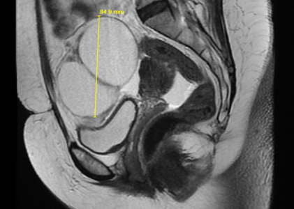
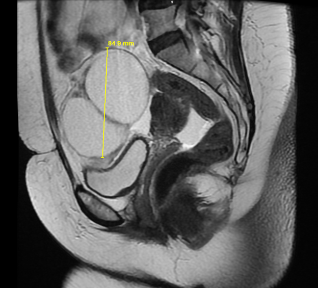
Ovaries: There is a complex multiseptated cystic lesion just to the right of midline in the anterior mid pelvis measuring 8.5 x 6.4 x 8.5 cm. This may arise from the right ovary. Neither ovary is otherwise seen.. No mural nodule is seen within the cystic lesion. The septations are thin and do not appear hypervascular. There is mild adjacent soft tissue edema. There is free fluid in the cul-de-sac.
Urinary Bladder: The urinary bladder appears normal, without stones or wall thickening.
Abdominal Wall: No abdominal wall or inguinal hernia is seen.
Lymphatics: No pelvic lymphadenopathy is present.
Bones: The bones appear unremarkable.
IMPRESSION: Multiseptated cystic lesion in the right pelvis, possibly arising from the right ovary. Findings are suspicious for ovarian malignancy; consider further evaluation with laparoscopy. There is a small amount of free fluid within the pelvis. No peritoneal nodule is seen.