


The following is our MRI Abdominal Enterography Case Study of the month. The patient came for exam for history of ulcerative duodenitis with worsening abdominal pain. The MRI Abdominal Enterography Case Study procedure included multi-planar images of the abdomen obtained without IV contrast on our 1.5 Tesla MRI machine. The comparison CT scan was done on 10/24/2015. For more information on MR Enterography visit this web page:
MRI Exam Findings:
MRI Abdomen Without IV Contrast
History: Enterography for ulcerative duodenitis with worsening abdominal pain
Comparison: CT 10/24/2015
Technique: Multiplanar images of the abdomen were obtained at 1.5 Tesla without IV contrast.
Findings:
Liver: The liver appears normal in size and contour. No mass or intrahepatic biliary dilatation is present. No areas of abnormal enhancement are seen.
Gallbladder: The gallbladder is contracted. No Evidence of gallstones or biliary dilatation.
CBD: The common bile duct is normal in caliber.
Pancreas: The pancreas appears normal.
Spleen: The spleen appears normal.
Adrenal Glands: The adrenal glands appear normal.
Kidneys: The kidneys appear normal. No mass or hydronephrosis is seen.
Aorta: No evidence of aortic aneurysm or dissection is seen.
Lymphatics: No abdominal lymphadenopathy is present.
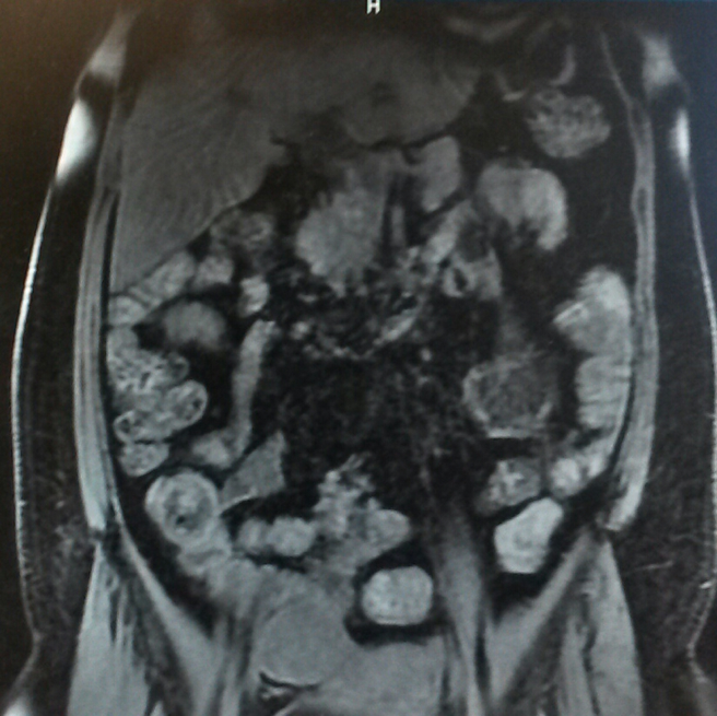 GI Tract: The patient has undergone prior small bowel resection in the right abdomen. There is no bowel obstruction. There is a moderate amount of air throughout the small bowel, but there is no localized bowel wall thickening.
Impression: No evidence of active inflammatory bowel disease. No localized bowel wall thickening or evidence of bowel obstruction.