


MRI Hand Case Study
Our case study of the month is an MRI scan of the right hand. The patient presented with soft tissue swelling. The MRI Hand Case Study procedure included coronal, axial, and sagittal images of the right hand with attention to the right second digit and they were obtained on our 1.5 Tesla MRI machine. There was no comparison scan.
MRI Exam Findings
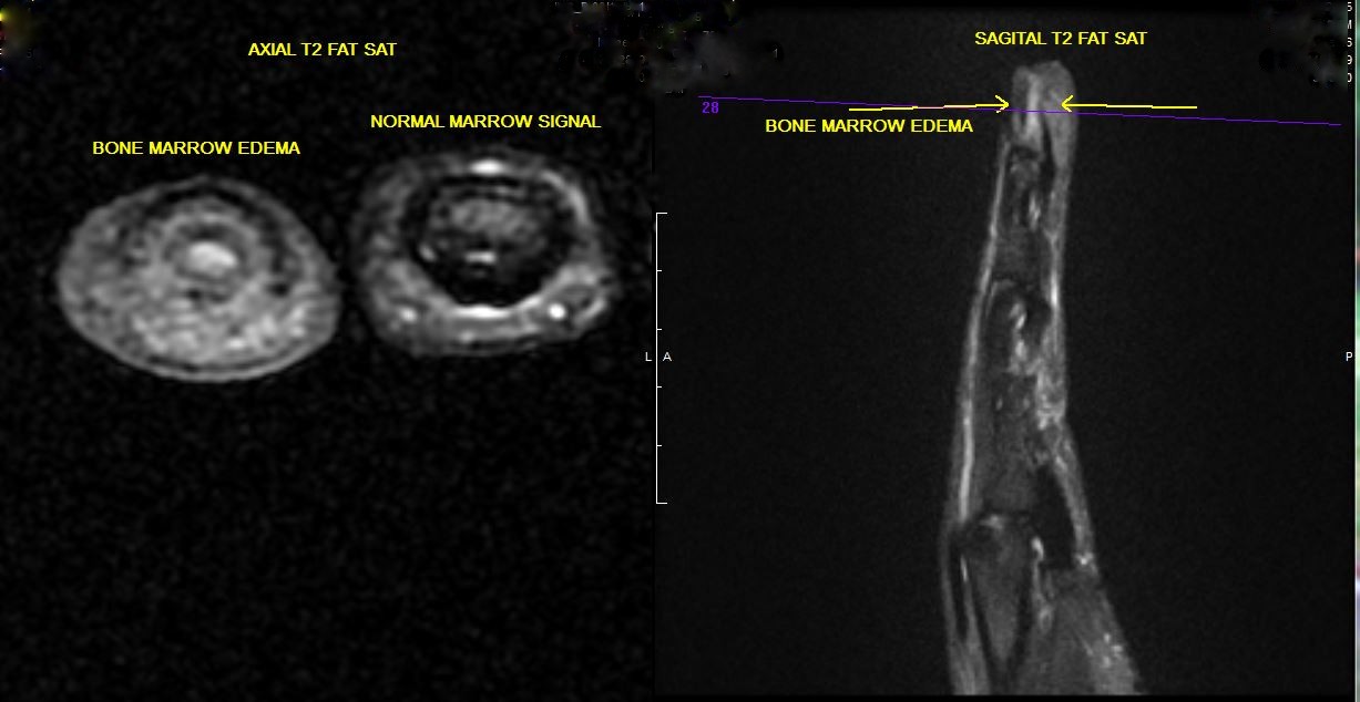
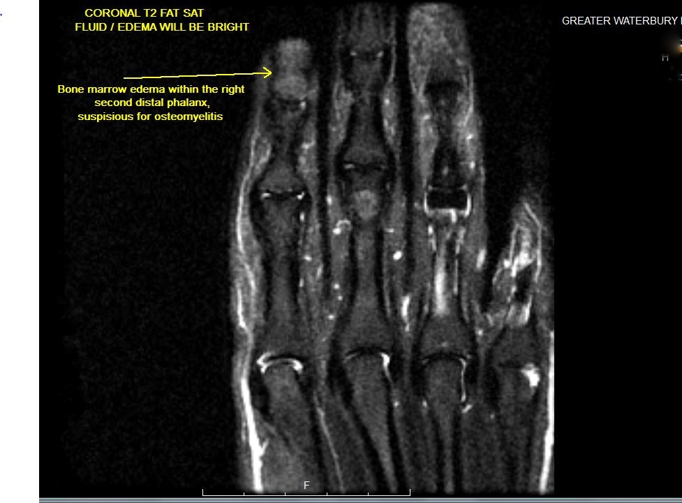
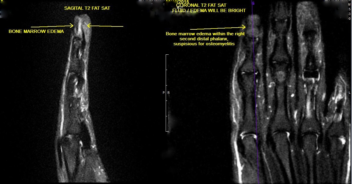
There is bone marrow edema involving the right second distal phalanx, most prominent at the level of the tuft of the distal phalanx, suspicious for osteomyelitis. There is a minor pattern of diffuse soft tissue swelling involving the distal aspect of the right second digit. No fluid collection is identified within the soft tissues.
MRI Exam Impressions
Bone marrow edema within the right second distal phalanx, suspicious for osteomyelitis.
For more information on MRI imaging services at Greater Waterbury Imaging Center, visit our clinical section of the website.