
Table of Contents
Vascular Malformations and Vasculitis
Blood vessels include arteries, veins, capillaries, and lymphatic vessels. Two broad categories of vascular conditions we encounter in body MRI are:
- Vascular malformations – developmental abnormalities of blood or lymph vessels. These are usually present from birth (congenital), though they may only become apparent later in life.
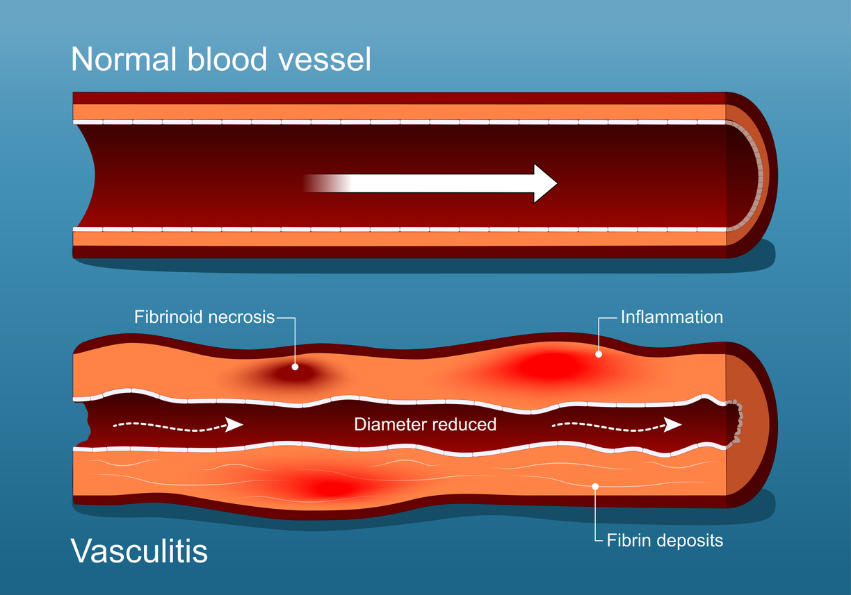
- Vasculitis (inflammation of the vessel wall) – conditions in which the wall of a blood vessel becomes inflamed, leading to damage, narrowing, or occlusion of the vessel.
Understanding the difference matters because MRI is often essential for accurate diagnosis, guiding treatment, and monitoring both vascular malformations and vasculitides.

How and Where They Occur
Vascular Malformations
These can involve one or more types of vessels (arteries, veins, capillaries, lymphatics) and may be classified by flow pattern (slow vs. fast) and vessel type. For example:
- Venous malformations (slow-flow) are among the most common malformations.
- Arteriovenous malformations (AVMs, fast-flow) – abnormal direct connections between arteries and veins, bypassing the capillary bed.
- Mixed or combined forms, e.g., venolymphatic malformations. They can occur anywhere in the body, including the limbs, trunk, head/neck, and internal organs. Because they are congenital, they may remain quiescent and only become symptomatic after triggers like puberty, pregnancy, trauma, or growth.
Vasculitis (Inflammation of Vessels)
Vasculitis may affect large, medium, or small vessels, depending on the disease. For example:
- Large-vessel vasculitis (LVV) such as Takayasu arteritis or Giant cell arteritis involve major arteries.
- The inflammation may lead to vessel wall thickening, narrowing (stenosis), aneurysm formation, or occlusion.
Imaging plays a key diagnostic role because symptoms may be nonspecific (e.g., fatigue, pain, ischemia) and mimic other conditions.
Why MRI Matters / What We Look For
MRI (and related techniques such as MR angiography (MRA) or vessel-wall imaging) is a powerful modality for both malformations and vasculitides.
Vascular Malformations
- MRI helps define the extent of abnormal vessels (arterial, venous, lymphatic), the flow character (slow vs. fast), and their relationship to adjacent tissues (muscle, bone, vital organs).
- Recognizing the flow type, such as fast-flow AVMs versus slow-flow venous malformations, on MRI is critical for planning treatments like embolization or surgery.
Vasculitis / Vessel Wall Inflammation
- High-resolution MRI vessel-wall imaging (VW-MRI) enables direct visualization of vessel wall thickening and contrast enhancement, which are key indicators of active vasculitis and inflammation.
- Imaging guides diagnosis, monitors response to therapy, and detects complications such as aneurysms or stenosis.
In short, for the body MRI service at Greater Waterbury Imaging Center, when a clinician or patient is concerned about either a vascular malformation or vasculitis, MRI is often one of the first advanced imaging steps.
Typical Signs, Symptoms & Why Referral Matters
Vascular Malformations
Symptoms vary by type and location:
- A visible birthmark or swelling may be present.
- Pain, swelling, and functional impairment (limb movement, joint bending) are associated with deep lesions.
- Internal malformations may manifest as bleeding, high‐output cardiac failure (in large AVMs), or compression of adjacent organs. Referral to a vascular anomaly specialist and advanced imaging is recommended, as management is often multimodal.
Vasculitis
Because vessel‐wall inflammation may affect many organs, the presentation is often wider:
- Constitutional symptoms (fever, fatigue), limbs or organs with reduced blood supply (claudication, stroke, renal involvement).
- Imaging suspicion may arise when lab signs of inflammation (ESR, CRP) are elevated, or when vascular imaging shows vessel narrowing or thickening. Early diagnosis is essential to prevent irreversible damage.
What Patients Can Expect at Our MRI Center
When referred for MRI to evaluate suspected malformation or vasculitis, the workflow typically includes:
- A clinical discussion with the ordering clinician and our radiologist to tailor the MRI protocol (for example: include contrast, include high-resolution vessel wall sequences, include MRA).
- MRI scan with the appropriate dedicated sequences (e.g., for malformations: flow studies, dynamic contrast, lesion mapping; for vasculitis: high-resolution T1 with contrast, black-blood sequences).
- Post-processing and radiologist interpretation, focusing on measuring lesion extent (for malformations) or vessel wall enhancement/thickening (for vasculitis).
- A detailed, clear radiology report delivered to the referring clinician (and patient as appropriate) with findings, implications, and often suggestions for follow-up or referral to a specialist.
- Our MRI services can establish a baseline for follow-up studies, helping monitor disease progression or response to therapy effectively.
At Greater Waterbury Imaging Center, we use high-quality MRI equipment and work closely with referring clinicians to ensure scans are optimized for vascular disorders, delivering both clarity and peace of mind to patients.
When is MRI Most Helpful – and When Not Enough?
MRI is beneficial, but there are limitations:
- Small vessels (especially in tiny vasculitides) may be below MRI resolution; additional modalities (ultrasound, PET/CT) may be required.
- For malformations involving rapid flow or complex microanatomy, catheter angiography (DSA) may remain the gold standard.
- MRI cannot always alone determine which malformations will progress or rupture; clinical context and specialist input remain critical.
Thus, MRI should be viewed as part of a comprehensive diagnostic program—not a stand-alone guarantee.
Take-home for Patients & Clinicians
- If you or your clinician suspects a vascular malformation (e.g., a swelling, birthmark, bleeding, or functional impairment) or vasculitis (e.g., unexplained systemic inflammation + vascular symptoms), consider an MRI early.
- At the same time, ensure your clinician discusses with the imaging centre specific protocols (contrast, flow sequences, vessel wall imaging) so that the MRI.
- Interpretation is best done in the context of a multidisciplinary team, including radiology, interventional radiology, vascular anomalies specialists, and rheumatology (for vasculitis).
- At Greater Waterbury Imaging Center, we aim to provide precise, timely imaging with reports that support clinical decision-making for both patients and referring clinicians.
Vascular malformations and vessel-wall inflammation (vasculitis) represent two essential categories of vascular disorders that can affect patients of all ages. Thanks to modern MRI techniques, imaging can now define the anatomy, flow characteristics, and vessel-wall changes, and guide therapy or monitoring.
Whether you’re a patient seeking clarity or a clinician seeking high-quality diagnostic imaging, our body MRI service at Greater Waterbury Imaging Center is equipped to help navigate these complex vascular conditions in a friendly, professional, and patient-centred manner.
References
- “Vascular Malformations: Symptoms, Treatment and Outlook.” Cleveland Clinic. Cleveland Clinic
- Schmidt VF et al., “Imaging of peripheral vascular malformations.” PMC. PMC
- “Vascular Malformations.” Johns Hopkins Medicine. Johns Hopkins Medicine
- Cox JA et al., “Vascular Malformations: A Review.” PMC. PMC
- Brahmbhatt AN et al., “Vascular lesions of the head and neck – Insights into Imaging.” Insights Imaging. SpringerOpen
- Popescu I et al., “Imaging in Large Vessel Vasculitis — A Narrative Review.” PMC. PMC
- Tawakol A et al., “Current and Emerging Approaches to Imaging Large Vessel Vasculitis.” Circ Imaging. AHA Journals
- Edjlali M et al., “Vessel Wall MR Imaging for the detection of intracranial …” AME Groups. Cardiovascular Diagnosis and Therapy
- Kang N et al., “Essentials for Interpreting Intracranial Vessel Wall MRI.” RSNA. RSNA Publications
- “EULAR recommendations for the use of imaging in large vessel vasculitis.” Ann Rheum Dis. Annals of the Rheumatic Diseases