

 Diseases of the Pancreas and Gallbladder (Using MRCP)
Table of Contents
- 1 Diseases of the Pancreas and Gallbladder (Using MRCP)
- 2 Understanding MRCP: A Noninvasive MRI Technique for Pancreatic and Biliary Imaging
- 3 When MRCP May Be Clinically Indicated
- 4 What to Expect During an MRCP Exam
- 5 Conditions Commonly Evaluated with MRCP
- 6 MRCP vs. Other Imaging Options
- 7 Safety and Limitations of MRCP
- 8 How MRCP Supports Fast, Accurate Diagnosis Without Invasive Procedures
- 9 More MRI Imaging Options for Liver, Kidneys, and Digestive Health
- 10 Greater Waterbury Imaging Center: Your Trusted MRI Provider
Understanding MRCP: A Noninvasive MRI Technique for Pancreatic and Biliary Imaging
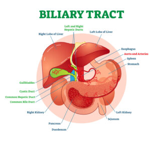
Magnetic Resonance Cholangiopancreatography (MRCP) is a specialized, noninvasive MRI technique that produces detailed images of the bile ducts, gallbladder, pancreas, and pancreatic ducts. Unlike traditional imaging methods that may involve endoscopy or ionizing radiation, MRCP uses magnetic fields and radio waves to visualize fluid-filled structures within the abdominal region.
By highlighting bile and pancreatic ducts in high contrast on T2-weighted MRI scans, MRCP enables radiologists to identify gallstones, tumors, inflammation, and structural abnormalities with greater clarity and precision. This makes it a safer alternative to Endoscopic Retrograde Cholangiopancreatography (ERCP), which requires sedation, carries a small risk of complications, and is generally used when therapeutic intervention is needed.
RadiologyInfo.org offers this quick video for more information: Your Radiologist Explains Magnetic Resonance Cholangiopancreatography (MRCP).
MRCP plays a crucial role in diagnosing conditions that may otherwise remain undetected or difficult to assess using ultrasound or CT alone. It is beneficial when symptoms such as abdominal pain, jaundice, or unexplained digestive issues suggest problems within the hepatobiliary or pancreatic systems.
When MRCP May Be Clinically Indicated

Your healthcare provider may recommend an MRCP scan when there is a need to examine the pancreas or biliary system more closely. It is instrumental in diagnosing or ruling out the following:
- Persistent or severe abdominal pain, especially when other tests are inconclusive.
- Gallstones or bile duct stones (choledocholithiasis), particularly when ultrasound does not provide clear results.
- Pancreatitis, to determine if stones, blockages, or ductal abnormalities are contributing to inflammation.
- Pancreatic or biliary tumors, including suspected malignancies or strictures in the ducts.
- Congenital anomalies such as pancreas divisum or pancreaticobiliary maljunction.
- Pre-surgical planning, especially when mapping the anatomy of biliary ducts, is crucial for procedures involving choledochal cysts or bile duct reconstruction.
By offering a detailed, radiation-free view of the hepatobiliary and pancreatic systems, MRCP enhances diagnostic accuracy while reducing patient risk.
What to Expect During an MRCP Exam
MRCP is typically performed as part of a standard MRI of the abdomen, with minimal preparation and no sedation in most cases.
Before the Exam
Patients are generally asked to fast for at least 4 hours prior to the procedure to reduce the amount of fluid and gas in the stomach and intestines, which can interfere with imaging quality. All metal objects, such as jewelry, glasses, and belts, should be removed to ensure safety in the MRI environment.
During the Exam
You will lie down on the MRI table, which slides into the scanner. The exam typically takes 10 to 30 minutes. MRCP uses high-powered magnets and radio waves to produce detailed images—no X-rays or invasive scopes are required. Most MRCP exams do not require contrast, though in some cases, gadolinium-based contrast agents may be used for better visualization of surrounding organs.
After the Exam
There is no recovery time needed unless sedation was required (which is rare for MRCP). A radiologist will review your images and send a detailed report to your referring provider, who will then discuss next steps with you based on the results.
Conditions Commonly Evaluated with MRCP
MRCP provides clear and detailed imaging of the biliary and pancreatic ducts, enabling radiologists and physicians to diagnose a wide range of conditions that may cause abdominal pain, jaundice, or digestive symptoms. Below are some of the most common disorders MRCP helps to identify.
Gallstones and Bile Duct Stones (Choledocholithiasis)
 MRCP is highly effective at detecting stones lodged in the bile ducts, even when ultrasound results are inconclusive. These stones can block bile flow and cause significant pain, nausea, or jaundice. If left untreated, choledocholithiasis can lead to severe complications, including cholangitis (infection of the bile ducts) or pancreatitis.
MRCP excels at identifying stones lodged in the bile ducts, even when standard tests, such as ultrasound and laboratory results, appear normal. A study published in the National Library of Medicine found MRCP to be a highly reliable method for detecting common bile duct stones, significantly reducing rates of misdiagnosis when biochemical indicators and ultrasound fail to indicate retained stones.
Pancreatitis

For patients with suspected pancreatitis, MRCP helps identify underlying causes such as gallstones, duct blockages, or pancreatic duct abnormalities. It’s beneficial for detecting biliary pancreatitis, which accounts for approximately 40% of acute pancreatitis cases. MRCP also assists in evaluating chronic pancreatitis by revealing ductal strictures, dilations, and potential calcifications.
Pancreatic and Biliary Tumors
MRCP provides a noninvasive way to visualize tumors, strictures, or masses within the pancreatic or bile ducts. When cancer is suspected, MRCP contributes to staging by mapping ductal anatomy, identifying obstruction, and supporting surgical or oncologic planning. While MRCP does not replace biopsy or contrast-enhanced studies for full staging, it serves as an essential first step in many evaluations.
Congenital Anomalies and Anatomic Variants
Certain congenital disorders may contribute to chronic abdominal symptoms or elevate cancer risk. MRCP is particularly helpful in diagnosing:
- Pancreas divisum: A condition where the pancreatic ducts fail to fuse, occurring in about 10% of individuals. It may cause recurrent pancreatitis.
- Pancreaticobiliary maljunction (PBM): A rare anomaly where the bile and pancreatic ducts join outside the duodenal wall, increasing the risk for inflammation and cancer.
- Choledochal cysts: These fluid-filled sacs in the bile ducts are best visualized with MRCP, which helps surgeons understand the cyst’s size, shape, and relation to surrounding structures.
MRCP vs. Other Imaging Options
MRCP Compared to Other Imaging Methods
MRCP is often the preferred diagnostic tool for evaluating the pancreas, gallbladder, and biliary tree because it offers detailed anatomical imaging with fewer risks associated with other procedures. Below is a comparison of MRCP with commonly used alternatives:
MRCP vs. ERCP
Endoscopic Retrograde Cholangiopancreatography (ERCP) is both diagnostic and therapeutic, but is invasive, requiring endoscopic access to the bile ducts. ERCP carries risks of complications such as pancreatitis, infections, and bleeding. In contrast, MRCP is noninvasive, does not require sedation, and eliminates the risk of procedural complications while still providing clear visualization of ductal anatomy.
MRCP vs. Ultrasound
Ultrasound is widely available and helpful in evaluating gallbladder disease, but it can miss small stones or subtle ductal abnormalities—especially in patients with obesity or excessive bowel gas. MRCP offers superior resolution for fluid-filled structures and multiplanar imaging capabilities, making it more reliable for comprehensive ductal evaluation.
MRCP vs. CT Scan
CT imaging is practical for identifying masses and inflammation, but involves exposure to ionizing radiation and may require contrast agents. MRCP avoids radiation and is more effective in visualizing bile and pancreatic ducts without the use of iodinated contrast, which can pose risks in patients with kidney impairment.
Comprehensive Diagnostic Value
MRCP allows for simultaneous evaluation of the liver, biliary tree, pancreas, and adjacent soft tissues. This makes it particularly useful for preoperative planning and assessing conditions with overlapping symptoms or uncertain causes.
By offering a non-invasive, highly detailed alternative to traditional imaging methods, MRCP is a valuable first-line tool for diagnosing pancreatobiliary diseases.
Safety and Limitations of MRCP
MRCP is generally safe and well-tolerated by most patients. It does not use radiation, and contrast agents are typically not required.
Safety Considerations
- Implants: Patients with pacemakers, cochlear implants, or certain metallic implants should notify their provider, as these may interfere with the MRI.
- Kidney disease: If contrast is used (rarely for MRCP alone), patients with impaired kidney function may require evaluation to avoid complications from gadolinium.
- Pregnancy: MRI is typically safe in pregnancy, but the decision to proceed with MRCP should be based on clinical need and discussed with your healthcare provider.
Limitations
- Diagnostic only: MRCP cannot be used to treat conditions; unlike ERCP, it cannot remove stones or place stents.
- Small lesions: Tiny stones or early mucosal abnormalities may occasionally be missed.
- Motion artifacts: Patients must remain still during the exam, as movement can blur images and reduce diagnostic quality.
Despite these limitations, MRCP remains one of the most informative and patient-friendly imaging tools for biliary and pancreatic disease.
How MRCP Supports Fast, Accurate Diagnosis Without Invasive Procedures
If you’re experiencing abdominal discomfort, jaundice, or digestive issues, MRCP can provide critical answers quickly and without unnecessary risk. Unlike more invasive procedures, MRCP enables your care team to visualize complex ductal anatomy with precision, without the need for sedation, endoscopy, or radiation. Whether your physician suspects stones, inflammation, or structural anomalies, MRCP offers a comprehensive view to guide treatment with clarity and confidence.
For clinicians, MRCP is a powerful diagnostic tool that enhances decision-making and surgical planning. It offers high-resolution, multiplanar imaging that highlights fluid-filled ducts and adjacent soft tissues, enabling faster and more accurate assessments of biliary and pancreatic disease. This efficiency can improve patient outcomes, avoid unnecessary procedures, and streamline referrals to specialists.
More MRI Imaging Options for Liver, Kidneys, and Digestive Health
To better understand how MRI supports abdominal imaging, visit our other Body MRI pages, such as:
If you or your provider believe MRCP may be the proper test for your symptoms or diagnosis, contact Greater Waterbury Imaging Center for more information or to schedule an appointment.
Greater Waterbury Imaging Center: Your Trusted MRI Provider
At Greater Waterbury Imaging Center, we understand the importance of receiving clear answers when facing abdominal discomfort or a suspected digestive disease. Our advanced MRI technology, including MRCP capabilities, enables noninvasive evaluation of the pancreas, gallbladder, bile ducts, and related structures with precision and safety. Whether you’re managing chronic symptoms or preparing for surgery, our experienced radiology team delivers reliable, high-resolution imaging to support timely and confident clinical decisions.
Greater Waterbury Imaging Center offers high-quality MRI services, prioritizing patient comfort, clinical accuracy, and prompt results. Our wide-bore scanner provides a more open and comfortable experience, invaluable for individuals with anxiety or claustrophobia. Contact us today to learn more or schedule your MRI for pancreatic, gallbladder, or biliary evaluation using MRCP.