Table of Contents
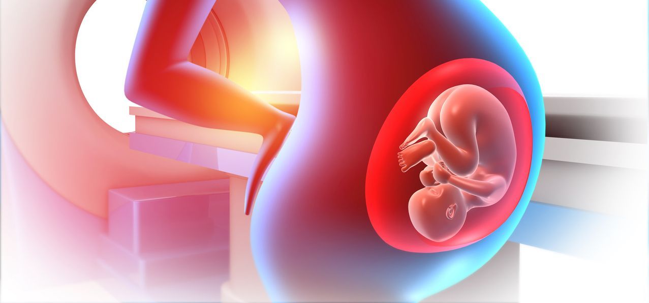
MRI During Pregnancy: Conditions MRI Can Help Assess
Pregnancy brings rapid changes in the body, and some symptoms require quick answers to ensure the safety of the parent and the baby. If ultrasound is unclear or more detail is needed, MRI is an effective option. MRI provides detailed images of soft tissues, organs, and the placenta without ionizing radiation. According to the Radiological Society of North America (1), both MRI (at ≤3T) and ultrasound are considered safe for routine use during pregnancy.
This page summarizes conditions, both related to and unrelated to pregnancy, for which MRI assists in evaluation, diagnosis, and care, complementing Greater Waterbury Imaging Center’s main Body MRI “Pregnancy” section.
Why MRI Is Used During Pregnancy
MRI is often used when ultrasound results are unclear or precise anatomy is needed for treatment. It helps:
- Diagnose urgent cases without radiation (e.g., acute abdominal pain).
- Identify masses or abnormalities seen on ultrasound (maternal or fetal).
- Assess placental issues when more details may affect delivery plans.
Safety Considerations: What Pregnant Patients and Clinicians Should Know
MRI does not use ionizing radiation
Unlike CT or X-rays, MRI does not expose the fetus to ionizing radiation. In normal clinical use, MRI is considered safe for the fetus.
First-trimester decision-making
Studies and safety guidelines show that first-trimester MRI does not increase risk to the fetus or in early childhood, but imaging should be based on clinical need. (2)
Gadolinium contrast is generally avoided in pregnancy
Most MRI scans during pregnancy are done without contrast. Gadolinium agents cross the placenta, so they should only be used when clearly needed, and the benefits outweigh potential fetal risks. (3)
Positioning and comfort matter
As pregnancy progresses, staying comfortable gets harder. Guidance suggests using a left-lateral offset in later stages to improve comfort and tolerance. (4)
Common Maternal Conditions Evaluated with MRI During Pregnancy
Acute abdominal pain and suspected appendicitis
Appendicitis serves as an illustrative case for the clinical utility of MRI; although ultrasound is typically the initial imaging modality, MRI is often employed when ultrasound findings are inconclusive. This approach facilitates timely diagnosis without radiation exposure. Comparative studies have assessed the performance of ultrasound versus MRI in pregnant patients with suspected appendicitis, highlighting the diagnostic capabilities of MRI in this context. (5)
Hepatobiliary disease (gallbladder, bile ducts, pancreas)
MRCP (MR cholangiopancreatography) noninvasively images the biliary tree and pancreatic ducts, making it helpful for pregnancy-related biliary symptoms—especially if ultrasound cannot clearly identify the problem or duct anatomy. (6)
Urinary tract obstruction and renal colic/hydronephrosis
Pregnancy may cause physiologic hydronephrosis, making it challenging to distinguish from obstruction on ultrasound. MRI (usually without contrast) can further assess the urinary tract and nearby tissues when needed. (7)
Gynecologic causes of pain: adnexal masses and suspected torsion
Adnexal masses are sometimes identified incidentally during obstetric ultrasound examinations or during assessments for pain. When ultrasound findings are inconclusive regarding the nature of an adnexal lesion, the superior soft-tissue resolution of MRI can enhance lesion characterization and assist in determining appropriate management strategies. (8)
Maternal masses and select cancer evaluations
While cancer evaluations are often adapted during pregnancy, MRI can help clarify the size and location of specific masses like those in the abdomen or pelvis, typically done without using contrast, when it might affect treatment or delivery plans. If contrast is considered, a thorough risk-benefit analysis is necessary beforehand. (9)
Placental and Uterine Conditions Where MRI May Help
MRI is used for further characterization in cases of placental disease or other obstetric complications that affect delivery planning, especially when ultrasound is limited by placental location or more anatomic detail is needed. (8)
Placenta-focused MRI helps clinicians plan safer deliveries when ultrasound cannot provide adequate information.
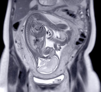
Fetal MRI: When Ultrasound Needs a Second Look
Fetal MRI supplements prenatal ultrasound by providing more explicit images of fetal anatomy and by further evaluating any abnormalities detected. There is no evidence that MRI harms the fetus, but IV contrast is avoided during pregnancy. (10)
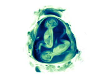
When is fetal MRI performed?
- According to practice guidelines, performing fetal MRI before 18 weeks of gestation typically does not provide additional information compared to high-quality (often transvaginal) ultrasound imaging. MRI becomes progressively more valuable later in pregnancy, as many anatomical structures can be visualized with greater detail at advanced gestational ages. (4)
- In clinical practice, fetal MRI is most frequently performed during the second or third trimester to evaluate further findings identified on ultrasound, except when an earlier examination is urgently indicated for medical reasons. (10)
What fetal conditions are commonly evaluated?
Fetal MRI is frequently used to assess abnormalities affecting the brain, spine, chest and lungs, abdomen and pelvis, and the placenta. It serves a vital role in supporting diagnosis, informing therapeutic discussions, and facilitating delivery planning. (10)
What to Expect During an MRI in Pregnancy
Most pregnancy MRI exams are outpatient and typically last less than an hour. Fetal MRIs usually take 30–50 minutes, using quick sequences to limit motion. You’ll be screened for metal and positioned comfortably. If you have claustrophobia or anxiety, inform your provider and imaging team ahead of time for support.
Imaging Access in Waterbury, CT
Greater Waterbury Imaging Center (GWIC) provides complete MR imaging services, (11) including options for claustrophobic patients. Located next to the Waterbury Hospital Emergency Department, GWIC offers private gated parking for convenience.
References
- Radiological Society of North America (RSNA). RSNA Statement on Safety of the Developing Fetus in Medical Imaging During Pregnancy (Reviewed 4/3/2018). https://www.rsna.org/uploadedfiles/rsna/content/role_based_pages/media/rsna-imaging-during-pregnancy-statement.pdf RSNA
- UCSF Radiology. CT and MR Pregnancy Guidelines (Patient Safety). https://radiology.ucsf.edu/patient-care/patient-safety/ct-mri-pregnancy UCSF Radiology
- American College of Radiology (ACR). Gadolinium Pregnancy Screening Statement (Aug. 23, 2022). https://www.acr.org/Advocacy/Position-Statements/Gadolinium-Pregnancy-Screening American College of Radiology
- ISUOG. Practice Guidelines (updated): performance of fetal magnetic resonance imaging (Ultrasound Obstet Gynecol, 2023). https://www.isuog.org/static/60706d51-b90e-4f9b-80a9008220319e68/Updated-ISUOG-Practice-Guidelines-performance-of-fetal-magnetic-resonance-1.pdf isuog.org
- Israel GM, Malguria N, McCarthy S, Copel J, Weinreb J. MRI vs. ultrasound for suspected appendicitis during pregnancy (PubMed abstract). https://pubmed.ncbi.nlm.nih.gov/18666160/ PubMed
- RadiologyInfo.org (RSNA/ACR). MRI Safety During Pregnancy (Reviewed May 1, 2023; PDF export). https://www.radiologyinfo.org/en/info/safety-mri-pregnancy?PdfExport=1 Radiologyinfo.org
- HSOA Journal Reproductive Medicine, Gynecology and Obstetrics, https://www.heraldopenaccess.us/article_pdf/63/mr-imaging-of-abdominal-pain-in-pregnant-women-a-review-of-common-obstetric-and-non-obstetric-pathologies.pdf
- Bonito G, Masselli G, et al. Imaging of Acute Abdominopelvic Pain in Pregnancy and Puerperium—Part I: Obstetric (Non-Fetal) Complications (Diagnostics, 2023). https://www.mdpi.com/2075-4418/13/18/2890 MDPI
- Gadolinium Pregnancy Screening Statement, https://www.acr.org/Advocacy/Position-Statements/Gadolinium-Pregnancy-Screening
- RadiologyInfo.org. Fetal MRI (Reviewed March 20, 2023). https://www.radiologyinfo.org/en/info/fetal-mri Radiologyinfo.org
- Greater Waterbury Imaging Center, https://greaterwaterburyimagingcenter.org/magnetic-resonance-imaging/