

Magnetic Resonance Imaging (MRI) is often recommended to help detect, diagnose, and monitor tumors in the chest, abdomen, or pelvis. MRI provides detailed images that assist physicians in evaluating these areas with accuracy and without exposing patients to radiation. Contact Greater Waterbury Imaging Center to schedule your MRI today.
General Overview
 A tumor is an abnormal growth of cells that can occur in nearly any organ or tissue. Tumors may be benign (non-cancerous) or malignant (cancerous). Benign tumors generally grow more slowly and remain localized, while malignant tumors may invade nearby tissues and spread (metastasize) to other areas of the body.
Tumors of the chest, abdomen, and pelvis can involve vital organs, including the lungs, liver, kidneys, pancreas, reproductive organs, or urinary bladder. Because these organs perform essential functions, accurate diagnosis and monitoring are critical to developing the right treatment plan.
MRI is a valuable imaging tool that provides exceptional soft tissue detail. It helps distinguish between benign and malignant lesions, guides treatment planning, and monitors outcomes after therapy.
MRI for Tumors of the Chest, Abdomen, or Pelvis
MRI is often chosen because of its ability to generate high-resolution, multiplanar images of soft tissues. Unlike CT scans, MRI does not use ionizing radiation, which can be especially important for patients requiring repeated follow-up imaging.
In clinical practice, MRI may be recommended when:
– A lesion is discovered on ultrasound or CT and requires further evaluation.
– A patient is undergoing treatment, and physicians need to assess the response.
– Staging is necessary to understand whether a tumor has spread locally or to nearby lymph nodes.
– Non-invasive assessment of vascular involvement is needed.
Compared to other modalities, MRI offers superior contrast resolution for many tumors. For example, it can better differentiate liver lesions, characterize pelvic masses, and evaluate mediastinal or chest wall tumors. In some cases, MRI is used in conjunction with CT, PET, or ultrasound to provide a comprehensive diagnostic picture.
Types of Tumors in the Chest, Abdomen, or Pelvis
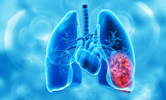
Chest
– Lung lesions: While CT is the primary tool for lung nodules, MRI can be used to evaluate chest wall invasion or large masses.
– Mediastinal tumors: MRI is helpful for thymomas, lymphomas, and neurogenic tumors.
– Chest wall and pleural masses: MRI can identify tumor extent and soft tissue involvement.
Abdomen
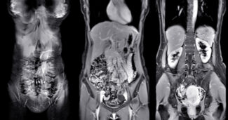 – Liver tumors: MRI plays a central role in diagnosing hepatocellular carcinoma, liver metastases, and benign lesions such as hemangiomas.
– Kidney tumors: Renal cell carcinoma, complex cystic lesions, and angiomyolipomas can be characterized with MRI.
– Adrenal masses: MRI helps differentiate adenomas from malignant lesions or pheochromocytomas.
– Pancreatic tumors: MRI and MRCP provide detailed visualization of the pancreas and bile ducts.
– Spleen and gastrointestinal tract: Though less common, tumors in these areas can also be evaluated with MRI.
Pelvis
 – Ovarian tumors: MRI can help distinguish benign cysts from malignant ovarian masses.
– Uterine tumors: Fibroids, endometrial cancers, and sarcomas can be characterized by MRI.
– Prostate cancer: Multiparametric MRI is now a key tool for prostate cancer detection and staging.
– Bladder tumors: MRI assists in evaluating tumor depth and muscle involvement.
– Soft tissue sarcomas: MRI provides excellent delineation of tumor size and relationship to surrounding structures.
How MRI Helps Evaluate Tumors
MRI is useful across the entire continuum of care:
– Detection: Identifies abnormal growths in patients with symptoms or incidental findings on other imaging tests.
– Characterization: Provides details about the nature of a mass—solid or cystic, vascular or non-vascular, benign or malignant appearance.
– Staging: Defines the size of the tumor, involvement of nearby tissues, and spread to lymph nodes or adjacent organs.
– Monitoring: Tracks changes over time, such as tumor shrinkage after chemotherapy or recurrence following surgery.
Additionally, MRI is often used to guide biopsies and surgical planning by providing precise maps of tumor location and extent.
What Patients Should Expect
An MRI exam is a safe, non-invasive procedure. Patients may receive contrast dye through an IV to improve visualization of certain tumors. Exams typically last 30 to 60 minutes, depending on the body area being studied.
For more information about how to prepare for an MRI, please visit our MRI Preparation Page: https://greaterwaterburyimagingcenter.org/magnetic-resonance-imaging/
Risks and Limitations
MRI is considered very safe for most patients. However, as with any procedure, there are some considerations:
– Contrast use: Rare allergic reactions or kidney concerns may occur.
– Claustrophobia: Some patients may feel anxious in the MRI scanner, but sedation options are available.
– Metal implants: Patients with certain pacemakers or devices may not be able to undergo MRI.
– Limitations: Very small lesions may be difficult to detect, and motion (such as breathing or bowel movement) can affect image quality.
Alternative or complementary imaging may include CT, PET, or ultrasound, depending on the clinical question.
References
- American College of Radiology. (2022). Management of Liver Cancer. ACR Appropriateness Criteria®., https://pubmed.ncbi.nlm.nih.gov/36436965/
- Nougaret, S., et al. (2019). MRI of tumors and tumor mimics in the female pelvis. Insights into Imaging, 10(1), Article 74. , https://pmc.ncbi.nlm.nih.gov/articles/PMC6677288/
- Abdominal Radiology Society (Expert Consensus). (2020). Use of pelvic MRI for staging of rectal cancer: Guidelines, https://abdominalradiology.org/
- Forstner, R., et al. (2016). Update on imaging of ovarian cancer. Current Radiology Reports, 4(1), 31. https://link.springer.com/article/10.1007/s40134-016-0157-9