

Lumbar Spine MRI Case Study
Our case study of the month is an lumbar spine MRI. The patient is a male with history of lumbar radiculitis. The MRI Lumbar Spine Case Study procedure included axial and sagittal images of the lumbar spine and they were obtained on our 1.5 Tesla MRI machine and were compared to a previous scan in 2013.
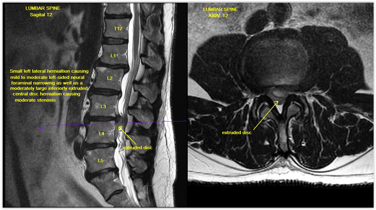
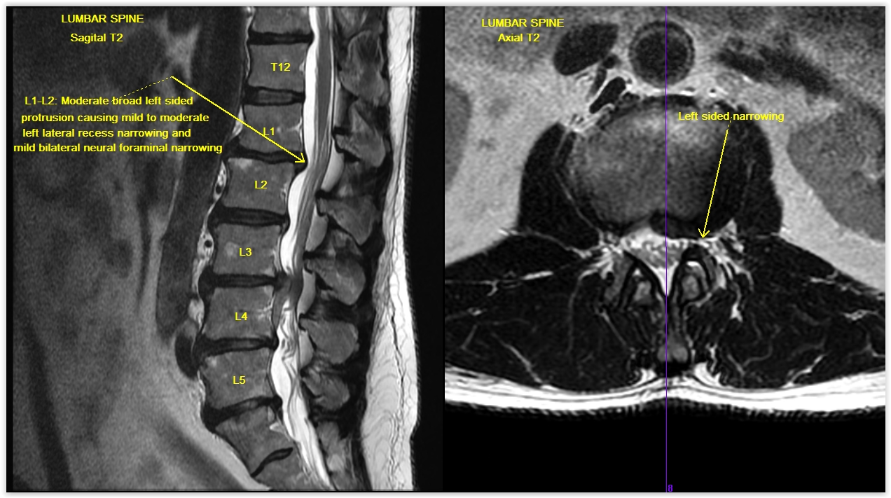
At L3-4, there is a moderately large inferiorly extruded disc herniation as well as a new small left lateral disc herniation at this same level, as discussed above. There is also multilevel degenerative disease with bulges and protrusions, similar to the previous examination, as discussed above from L1 through S1. A small protrusion at T11-12 is also noted.
Click here for more detail on this case study or to review how an MRI of the Lumbar Spine is done please visit our website page on MRI Lumbar Spine.