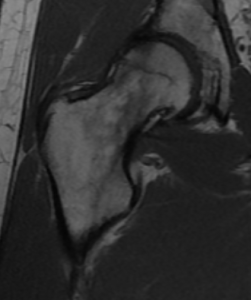
MRI RIGHT HIP
HISTORY: RIGHT HIP PAIN
COMPARISON: None
TECHNIQUE: Multiplanar images of the right hip were obtained at 1.5 Tesla without IV contrast.
FINDINGS:
BONES: No fracture or dislocation is seen. There are moderate degenerative changes in the right hip. The superior right hip joint space narrowing. Subchondral bone marrow edema in the superolateral right acetabulum and superior right femoral head and neck. There is no evidence of avascular necrosis.
MUSCLES AND TENDONS: There is normal signal in the visualized muscles and tendons.
ACETABULAR LABRUM: The labrum is grossly intact.
SOFT TISSUES: There is normal signal in the visualized soft tissues. No mass or abnormal fluid collection is seen.
IMPRESSION: Moderate right hip degenerative changes. No labral tear.


