CASE STUDY: MR RIGHT SHOULDER
HISTORY: SEVERE BILATERAL SHOULDER PAIN
COMPARISON: None
TECHNIQUE: Multiplanar images of the right shoulder were obtained at 1.5 Tesla without IV contrast
FINDINGS:
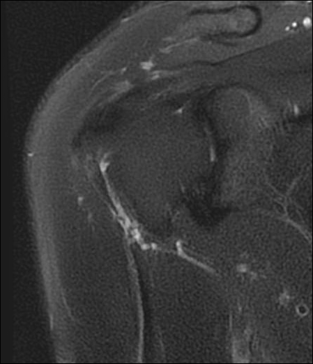
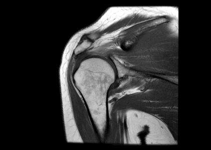
AC JOINT: There are mild hypertrophic degenerative changes of the acromioclavicular joint. There is mild narrowing of the subacromial space. There is minimal fluid within the subacromial subdeltoid bursa.
ROTATOR CUFF TENDONS:
Supraspinatus: There is thickening and edema of the distal supraspinatus tendon, with a partial interstitial tear of the tendon distally at its humeral insertion. There is reactive bone marrow edema within the greater tuberosity of the humerus.
Infraspinatus: There is abnormal signal along the distal infraspinatus tendon consistent with tendinosis.
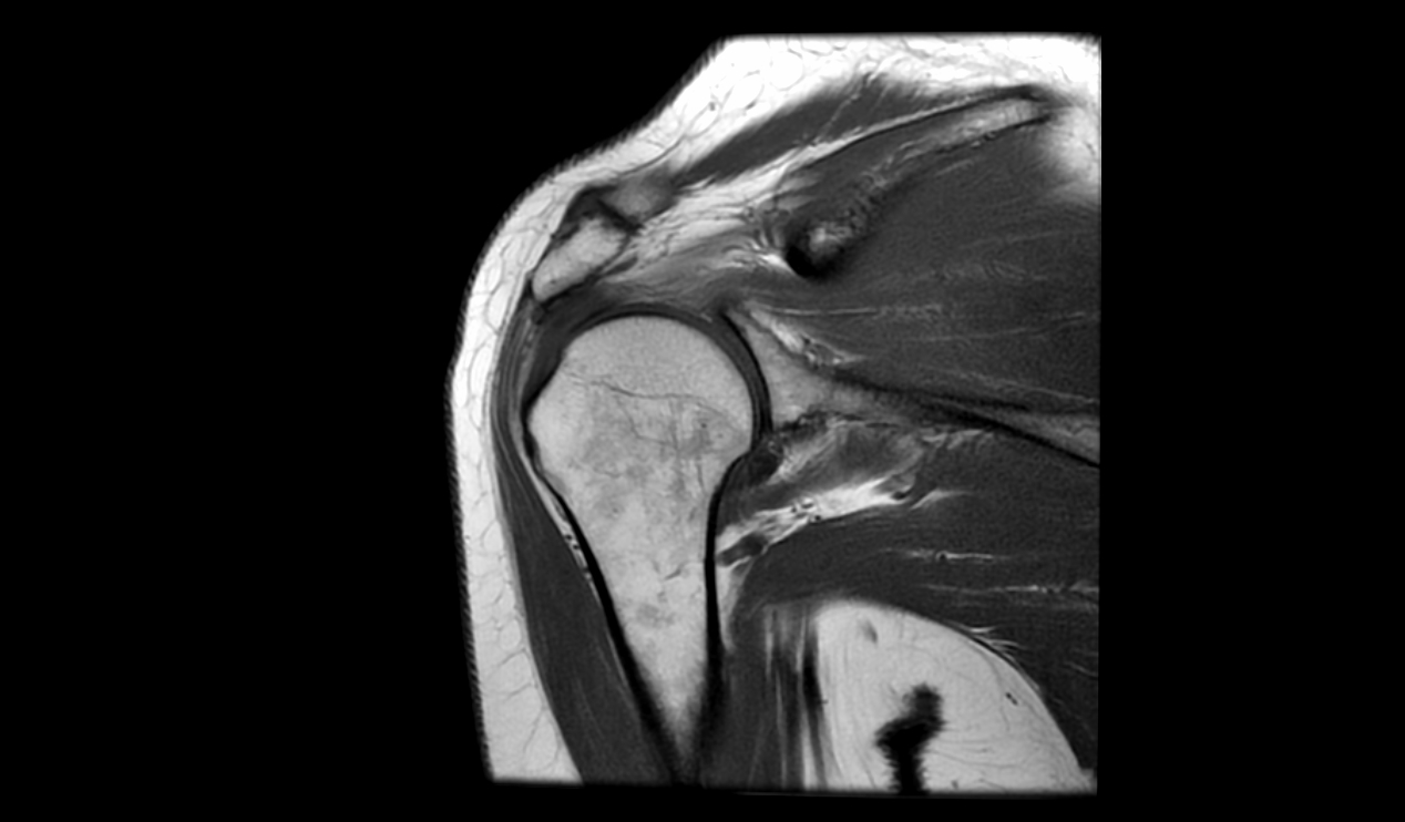
Subscapularis: The subscapularis tendon appears normal.
Teres Minor: The teres minor tendon appears normal.
GLENOHUMERAL JOINT: The glenohumeral joint appears normal.
BICEPS TENDON: The biceps tendon attachment is intact.
LABRIUM: The glenoid labrum appears intact.
IMPRESSION: Narrow subacromial space, with secondary degenerative changes in the rotator cuff tendons and a partial interstitial tear involving the distal supraspinatus tendon at its humeral insertion. No full-thickness rotator cuff tear.