MRI Female Abdominal Case Study with Pregnancy
Our case study of the month is an MRI scan of the abdomen without IV contrast. The patient is pregnant in late term. The MRI Female Abdominal Case Study procedure included multi-planar images of the abdomen without IV contrast and the scan was obtained on our 1.5 Tesla MRI machine. There was no comparison study for this patient.
MRI Exam Findings
History: Evaluated for Hernia
Technique: Multi-planar images of the abdomen were obtained at 1.5 Tesla without IV contrast.
Findings::
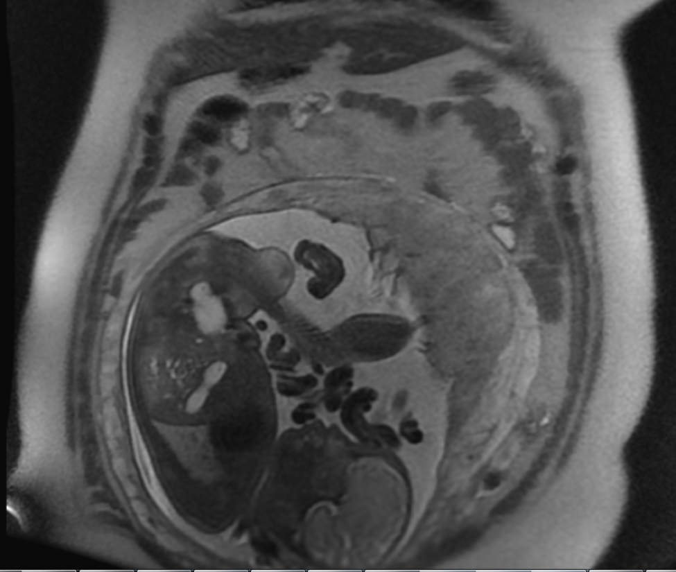
Pelvis: There is late term intrauterine gestation. The fetus is in the vertex lie. The placenta is along the left lateral and fundal walls. There is no fetal hydrocephalus or hydronephrosis. The appendix is not directly visualized, however no evidence of a distended, fluid-filled viscus in the right lower quadrant seen. There is no bowel wall thickening or adjacent inflammatory changes to suggest acute appendicitis. There is no bowel obstruction.
Liver: The liver is normal in size and contour. No mass or intrahepatic biliary dilatation is present.
Gallbladder: The gallbladder appears normal, without gallstones, wall thickening or pericholecystic fluid.
CBD: The common bile duct is normal in caliber. measuring approximately 2 mm.
Pancreas: The pancreas appears normal.
Spleen: The spleen appears normal.
Adrenal Glands: the adrenal glands appear normal.
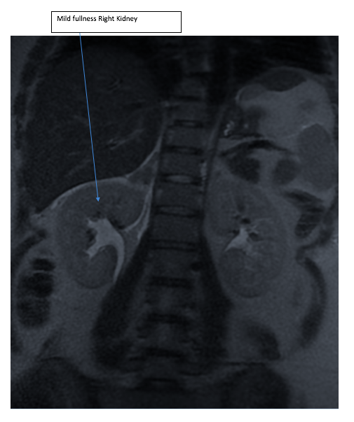
Kidneys: There is mild fullness of the right renal collecting system to the level of the uterus. The left kidney is normal.
Aorta: No evidence of aortic aneurysm or dissection is seen.
Lymphatics: No abdominal lymphadenopathy is present.
Impression: Levocurvature gestation. No evidence of acute appendicitis. Mild fulness right renal collection system. No gall stones or biliary obstruction.