

 MR PELVIS WITH AND WITHOUT IV CONTRAST:
HISTORY: ELEVATED PROSTATE-SPECIFIC ANTIGEN
COMPARISON: None.
TECHNIQUE: Multiplanar images of the pelvis were obtained at 1.5 Tesla prior to and following administration of 16 ml of Dotarem intravenous contrast. 3-D evaluation was performed on an independent workstation with concurrent supervision.
During this public health emergency, we are using enhanced sterilization processes, social distancing measures and PPE for your protection.
FINDINGS:
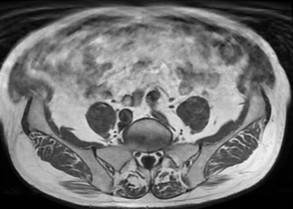
Urinary Bladder: The urinary bladder appears normal, without stones or wall thickening.
Prostate: The prostate measures 5.5 x 3.5 x 4.6 cm, corresponding to an estimated size of 46 g. Per precontrast images, there is heterogeneous signal throughout the central and transitional zones. There is heterogeneous signal along the posterior lateral peripheral zones. On diffusion-weighted images, there is an ovoid area of moderate diffusion restriction within the left anterolateral transitional zone in the mid-prostate body measuring up to 2.0 cm in length. The postcontrast images, this area is hypervascular. There is no evidence of extracapsular prostate cancer or neurovascular bundle encasement.
Seminal Vesicles: The seminal vesicals are symmetric bilaterally and appear unremarkable. The right testis is seen in the Iower right inguinal canal.
Lymphatics: No pelvic lymphadenopathy is present.
Bones: There is no fracture or bone lesion.”
Soft tissues: There is extensive sigmoid diverticulosis without evidence of acute diverticulitis.
IMPRESSION: 2.0 cm focus of abnormal diffusion restriction and hyperenhancement in the left anterolateral transitional zone in the mid-body of the prostate, moderately suspicious for malignancy. There is no evidence of extracapsular
prostate cancer or metastatic disease.
PIRADS 4: High (clinically significant cancer is likely to be present)