HISTORY: INTRAMURAL AND SUBSEROUS LEIOMYOMA
COMPARISON: CT 11/12/2013
TECHNIQUE: Multiplanar images of the pelvis were obtained at 1.5 Tesla prior to and following administration of 6.5 mL of Gadavist intravenous contrast.
FINDINGS:
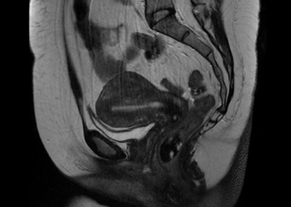
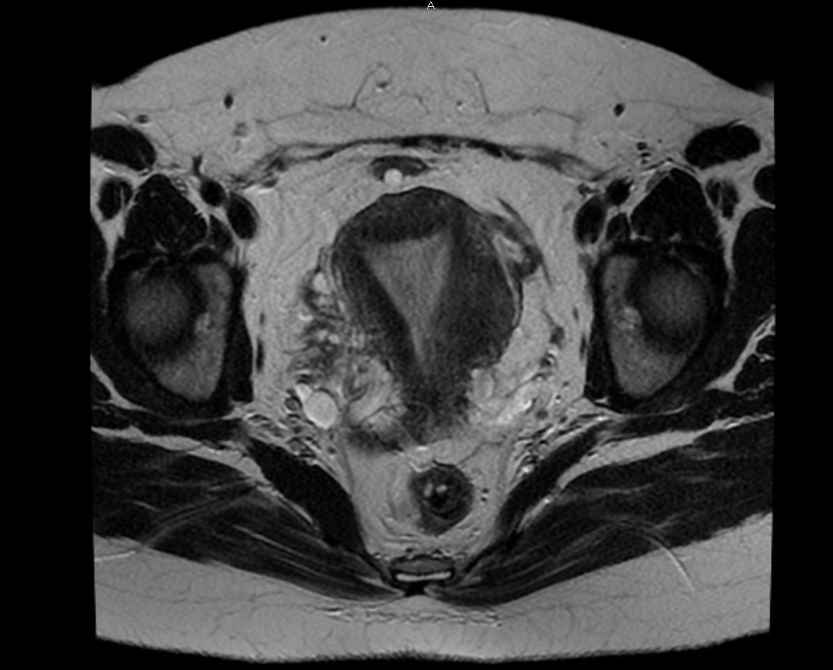
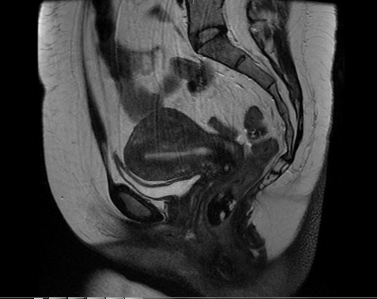
Uterus: The uterus is enlarged measuring 9.3 cm in length. There is a 2.9 cm intramural fibroid of the let posterior uterine fundus. There is diffuse thickening of the junctional zone of the uterus, suspicious for adenomyosis.
Endometrium: The endometrium is normal in thickness.
Ovaries: There are multiple tiny follicles scattered throughout both ovaries. No adnexal mass.
Urinary Bladder: The urinary bladder appears normal, without stones or wall thickening.
Abdominal Wall: No abdominal wall or inguinal hernia is seen.
Lymphatics: No pelvic lymphadenopathy is present.
Bones: The bones appear unremarkable.
IMPRESSION:
Enlarged fibroid uterus, with a 2.9 intramural fibroid of the left posterior fundus. Diffusely thickened junctional zone of the uterus, suspicious for adenomyosis. No other uterine mass. No adnexal mass.