


MRI Ankle Case Study
 Our case study of the month is an MRI scan of the left ankle, for an injury while playing basketball. The 18 year old male patient presented with left ankle pain. The MRI Ankle Case Study procedure included multi-planar images of the left ankle obtained without IV contrast on our 1.5 Tesla MRI machine. There was no comparison scan.
MRI Exam Findings:
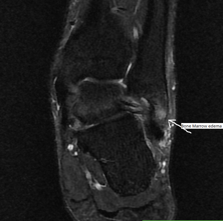
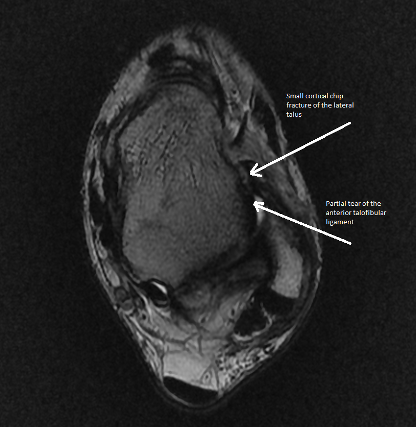 Bones: There is bone marrow edema involving the lateral malleolus, consistent with a bone contusion. A small cortical chip fracture involves the lateral aspect of the talus anteriorly, at the attachment of the anterior talofibular ligament.
Tendons: The flexor and extensor tendons are intact. The Achilles tendon and plantar fascia are intact.
Ligaments: There is a partial tear of the anterior talofibular ligament. The other ligaments appear to be intact medially and laterally.
Soft Tissues: No discrete soft tissue mass or focal fluid collection.
Impression: Partial tear of the anterior talofibular ligament, with a small cortical avulsion fracture involving the lateral aspect of the talus anteriorly at the attachment of the anterior talofibular ligament and a bone contusion of the lateral malleolus.
For more information on MRI imaging services at Greater Waterbury Imaging Center, visit our clinical section of the website.