
What Is an MRI-Guided Breast Biopsy?
An MRI-guided breast biopsy (also called an MR-guided breast biopsy) is a needle biopsy performed inside the MRI scanner. It is used when an area of concern is visible on a breast MRI and cannot be seen on a mammogram or ultrasound, so the MRI images are used to guide the needle to the exact spot. A small sample of tissue is removed and sent to a pathologist, who determines whether the area is benign or cancerous [2].
This page is for two readers. If your breast MRI report recommends a biopsy, it explains what will happen, why this type of biopsy was chosen, and what the results mean. If you are a referring physician or breast surgeon, it lays out the indications, the technique, and how to send a patient to Greater Waterbury Imaging Center.
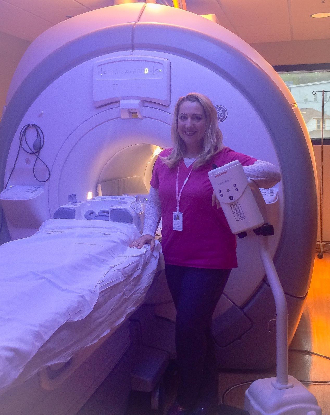
Greater Waterbury Imaging Center performs MRI-guided breast biopsy on site in Waterbury on referral from your physician, on the same wide-bore 1.5T scanner and dedicated breast coil used for breast MRI. The center holds ACR Breast MRI Accreditation, current through 2029. That matters for this procedure: the American College of Radiology expects a facility that performs breast MRI to have MRI-guided biopsy available, either on site or through a referral arrangement, so that a finding seen only on MRI can be sampled [4]. At GWIC it is available on site.
Why MRI Guidance, and Where It Fits Among the Other Biopsy Methods
The imaging method that found the lesion is the method that has to guide the needle. A radiologist can only sample what the guidance images show, so the choice of biopsy is set by which exam can see the area of concern [2].
Ultrasound-guided biopsy is used when the finding is visible on ultrasound. It is the quickest and most comfortable option, and most breast biopsies are done this way.
Stereotactic biopsy uses mammography to guide the needle. It is the standard method for calcifications and for masses seen on a mammogram.
MRI-guided biopsy is reserved for findings seen only on breast MRI. Breast MRI uses IV contrast to show blood flow, so it can reveal an area of abnormal enhancement that shows nothing on a mammogram or ultrasound. When a second look with ultrasound cannot locate the area, MRI is the only way to sample it [2, 3].
One limitation belongs up front. Some MRI findings take up contrast differently from day to day, and if the area cannot be seen on the biopsy day, the radiologist will cancel the procedure instead of sampling tissue that cannot be confirmed as the target. When that happens, follow-up MRI in a few months is the usual next step [3].
How the Procedure Works
Before the biopsy
Your physician or breast surgeon faxes the order to the center, and the center requests the breast MRI that showed the finding along with any other prior breast imaging, since the radiologist plans the biopsy from those images. Most patients referred for this biopsy had their breast MRI at Greater Waterbury Imaging Center, so those images are already on file.
When your appointment is booked, the scheduling team gives you the pre-procedure instructions. The center sets no medication requirements before the biopsy; eat normally and take your medications as usual. Do tell the scheduler what you take, including aspirin, blood thinners, and supplements, so the radiologist knows before the procedure, and never stop a prescription medication on your own. Some referring physicians choose to pause a blood thinner beforehand; if yours does, that instruction comes from your physician [2].
Because the procedure uses gadolinium contrast, you will be screened for kidney function, prior contrast reactions, and pregnancy, the same screening used for a diagnostic breast MRI. Tell the technologist about any implants, devices, or metal in your body; the MRI safety page covers what to report. The biopsy is done with local anesthetic only. Sedation is not used at the center, so you can drive yourself home.
During the biopsy
You lie face down on a padded table with the breast positioned through an opening into the breast coil, the same position used for a diagnostic breast MRI. An IV is placed in your arm for the contrast. The breast is gently held between two plates, one of which carries a grid that the radiologist uses to plan the needle path [2].
A first set of images is taken with contrast to confirm the target is visible. Using those images, the radiologist calculates the position and depth of the lesion, numbs the skin and deeper tissue with local anesthetic, and makes a small nick in the skin. The biopsy needle is advanced to the calculated position, and a second set of images confirms it is in the right place before any tissue is taken [2, 3].
Greater Waterbury Imaging Center performs vacuum-assisted MRI-guided biopsy, the standard technique for this procedure: gentle suction pulls tissue into the needle, and the needle rotates to collect several samples through one insertion, typically 8 to 12 [2]. You will hear clicks and feel pressure, and you should feel little pain. A tiny marker is placed at the biopsy site so the area can be found again on future imaging; it is MRI-safe and will not set off metal detectors [2]. The procedure takes about an hour. You are awake throughout, and the technologist stays in contact with you.
After the biopsy
The technologist applies pressure and a dressing. No stitches are needed. Bruising and mild soreness for a few days are normal, and an over-the-counter pain reliever and a cold pack are usually enough. Avoid strenuous activity for 24 hours, then return to your normal routine. Call your physician for heavy bleeding, increasing swelling, drainage, redness, or warmth at the site [2].
A mammogram is taken immediately after the biopsy to document the marker’s position [2]. Because Greater Waterbury Imaging Center is an MRI-only center, the hospital’s imaging center performs the mammogram, and staff will direct you there after the biopsy. Plan on about 30 minutes. Once the mammogram is done, you can go home.
When Your Doctor May Order an MRI-Guided Breast Biopsy
A suspicious finding seen only on breast MRI
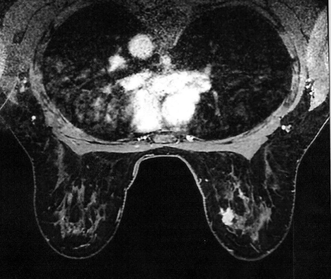
The most common reason is a breast MRI report that assigns the finding a BI-RADS category of 4 (suspicious) or 5 (highly suggestive of malignancy) and recommends tissue sampling. Category 4 covers a wide range of concern, so many category 4 biopsies come back benign; the biopsy is what settles the question [3]. If the finding could also be seen on ultrasound, the radiologist will usually try a targeted ultrasound first, because that biopsy is simpler. When ultrasound cannot find it, MRI guidance is the answer. Our Breast Cancer Awareness Month post explains every BI-RADS category and what each one means for your next step.
A second area found during staging of a known cancer
When breast MRI is used to map the extent of a diagnosed cancer, it sometimes shows an additional area of enhancement in the same breast or the opposite breast. The surgeon needs to know whether that area is cancer before planning surgery, because it can change the operation from a lumpectomy to a larger procedure. Our page on breast cancer detection and staging with breast MRI explains how MRI is used for staging.
A new finding on a high-risk screening MRI
Women at high lifetime risk of breast cancer, which the American Cancer Society defines as roughly 20 percent or greater by risk models, a known BRCA1 or BRCA2 variant, or chest radiation before age 30, are advised to have an annual breast MRI in addition to their mammogram, typically starting around age 30 [1]. Screening MRI finds cancers mammography misses, and it also finds benign areas that need sampling to prove they are benign. An MRI-only finding on a screening exam is a common route to this biopsy.
A finding that a second-look ultrasound could not locate
When an MRI finding is small, deep, or lacks a mass shape, ultrasound frequently cannot match it. The radiologist documents the attempt and proceeds to MRI guidance so the finding is sampled under the exam that can see it [2, 3].
For referring physicians and breast surgeons
Fax the order to the center with the breast MRI report that shows the target and the patient’s anticoagulation status; fax numbers and order forms are on our referring physician tools page. The center requests the prior breast imaging directly, and most patients referred for this biopsy had their breast MRI at GWIC, so the images are already on file. Note whether a targeted ultrasound was attempted, so the radiologist can plan MRI guidance without repeating that step. If the patient has a known malignancy, include the pathology and the planned surgery date. The center performs vacuum-assisted biopsy only; it does not offer MRI-guided wire or seed localization for surgery. The center schedules the procedure, gives the patient the pre-procedure instructions, sets no medication requirements of its own, and returns the pathology report and the concordance note to you.
What the Results Mean
The tissue goes to a pathologist, and the report returns to the radiologist and to your referring physician, who reviews it with you. Ask the center or your physician how long to expect; most results are available within several business days.
Before the result is final, the radiologist compares the pathology to the images, a step called radiologic-pathologic concordance. The question is whether the tissue result explains what the MRI showed. A benign result that explains the finding is reassuring, and follow-up imaging is typically recommended to confirm stability. A benign result that does not explain the finding is discordant, and the radiologist may recommend repeat biopsy or surgical excision to ensure nothing is missed [3].
Three outcomes are common. Benign findings such as fibroadenomas or fibrocystic change need no treatment, though your physician may schedule follow-up imaging. High-risk lesions such as atypical hyperplasia are not cancer but carry a higher chance that cancer is nearby, and surgical excision is often recommended. Malignant results go to your breast surgeon and oncology team, and the marker placed at biopsy helps them plan surgery [2, 3].
Benefits and Risks
MRI-guided biopsy samples a finding that no other method can reach, which is its entire purpose. For a patient, that means an MRI-only finding gets a definitive answer without surgery, and a surgeon planning an operation gets tissue proof before deciding how much to remove. Compared with a surgical biopsy, it uses a skin nick rather than an incision, leaves little or no scar, uses no radiation, takes about an hour, and has a recovery measured in days rather than weeks [2].
The risks are small and worth knowing. Bleeding into the breast (a hematoma) happens in under 1 percent of patients. Infection needing antibiotics happens in fewer than 1 in 1,000. There is a small chance the sample does not give a final answer, in which case surgical biopsy may follow. Deep lesions near the chest wall carry a very rare risk of the needle passing the chest wall [2].
Frequently Asked Questions About MRI-Guided Breast Biopsy
Most patients feel a pinch and a brief sting from the local anesthetic, then pressure during sampling. The breast itself should be numb. The most common complaint is lying face down for the length of the procedure, and cushioning helps.
Plan on about two hours in all. Arrive 30 minutes before your appointment for check-in and screening; the biopsy itself takes about an hour, and you then spend about 30 minutes at the hospital’s imaging center for the mammogram that confirms the marker position.
No. The biopsy is done with local anesthetic only, and sedation is not used at Greater Waterbury Imaging Center, so you can drive yourself home. If you would rather have company, you are welcome to bring someone.
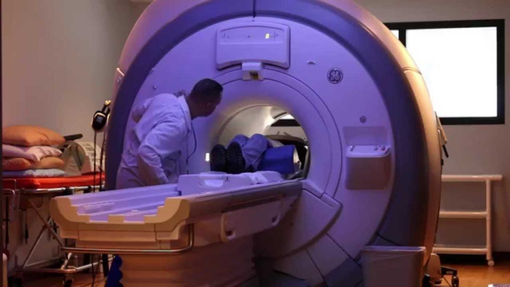
Yes. Greater Waterbury Imaging Center’s wide-bore 1.5T scanner has a larger opening than a traditional MRI, and you lie face down with your head turned to the side rather than looking up into the bore. Our claustrophobia page describes the options available before your appointment.
No. Greater Waterbury Imaging Center is an MRI-only center. Mammography, ultrasound, and biopsies guided by those exams are done at your breast imaging center. Your physician refers you to GWIC when the finding can only be seen on MRI.
A biopsy ordered for a suspicious finding is a diagnostic procedure, and most plans cover it subject to your deductible and coinsurance. Our office verifies benefits before your appointment and will tell you what to expect.
If Your Breast MRI Report Recommends a Biopsy
If your breast MRI report recommends a biopsy, or your breast surgeon needs an MRI-only finding sampled before surgery, Greater Waterbury Imaging Center performs MRI-guided breast biopsy in Waterbury on a wide-bore 1.5T scanner, on referral from your physician. Ask your doctor to fax the order to GWIC, or contact us, and we will coordinate the appointment with your physician’s office.
Greater Waterbury Imaging Center provides breast MRI and MRI-guided breast biopsy for patients throughout greater Waterbury, with evening and Saturday hours on a wide-bore 1.5T scanner. If you are anxious about the exam, see our claustrophobia page; for what to bring and how to prepare, see preparing for an MRI. Contact us to schedule an appointment or ask a question about your referral.
References
- American Cancer Society. American Cancer Society Recommendations for the Early Detection of Breast Cancer. Last revised July 23, 2026.
- Radiological Society of North America and American College of Radiology. MR-Guided Breast Biopsy. RadiologyInfo.org. Reviewed June 15, 2026.
- American College of Radiology. ACR Practice Parameter for the Performance of Magnetic Resonance Imaging-Guided Breast Interventional Procedures. Revised 2026 (Resolution 22).
- American College of Radiology. Breast MRI Accreditation Program requirements. As reported in ACR launches breast MRI program, AuntMinnie, April 8, 2010.