


Our case study of the month is an MRI scan of the left knee. The MRI Left Knee Case Study procedure included multi-planar images of the left knee obtained without IV contrast on our 1.5 Tesla MRI machine. There was no comparison scan.
Technique: Multiplanar images of the left knee were obtained at 1.5 Tesla without IV contrast.
Ligaments:
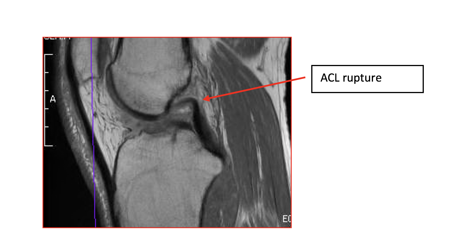 ACL: There is a complete ACL tear.
PCL: The posterior cruciate ligament is intact.
MCL: There is thickening of the deep fibers of the MCL suggesting a sprain and / or partial tear.
LCL: The lateral collateral ligament is normal.
Menisci:
Medial Meniscus: There is a questionable small tear of the posterior horn of the medial meniscus.
Lateral Meniscus: The lateral meniscus is intact.
Bones: There is moderate marrow edema consistent with bone bruising in the medial tibial metaphysis, most significant posteriorly with a small superimposed osteochondral defect posteriorly as well. No significant arthropathy is seen.
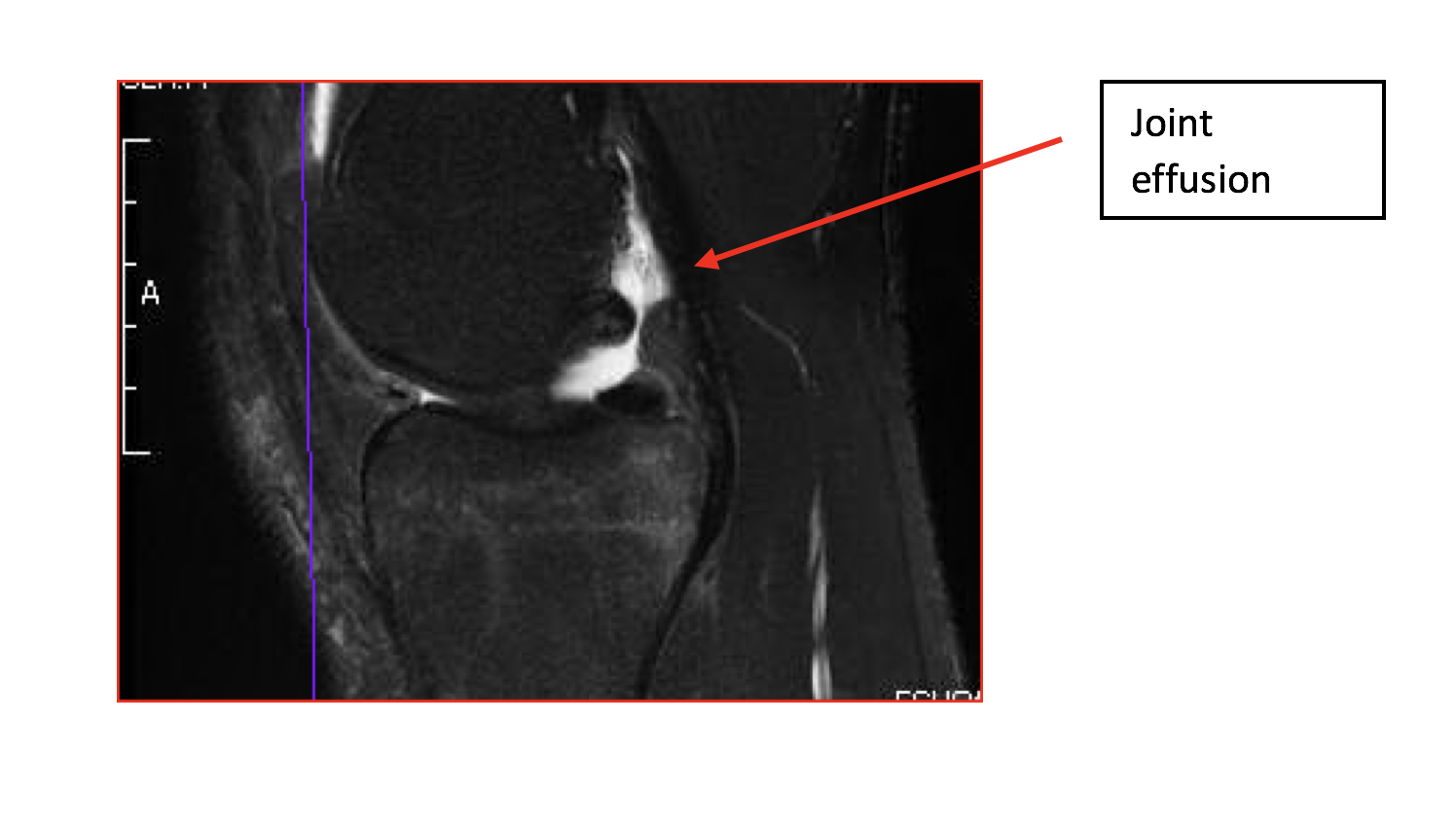
Soft Tissues: The quadriceps and patellar tendons are intact. There is scattered soft tissue edema. There is a moderate-sized joint effusion.
 Impression: Complete ACL tear. Thickening of the deep fibers of the MCL suggesting a sprain and / or partial tear. Questionable small medial meniscal tear. Findings consistent with bone bruising of the medial tibial metaphysis with small superimposed osteochondral defect, as discussed above. Moderate-sized joint effusion.