HISTORY: RIGHT LOBE MASS, HISTORY OF CIRRHOSIS
COMPARISON: Abdominal ultrasound 6/22/12
TECHNIQUE: Multiplanar images of the abdomen were obtained at 1.5 Tesla prior to and following administration of IV contrast.
FINDINGS:
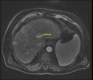
Liver: The liver is diffusely heterogeneous nodular in contour consistent with cirrhosis. There are no enhancing liver masses seen. There is a nonenhancing 10 mm lesion within the medial segment of the left lobe of the liver probably represents a cyst. The hepatic and portal veins are patent.
Gallbladder: Multiple small gallstones are present. There is no gallbladder wall thickening or pericholecystic fluid.
CBD: The common bile duct is normal in caliber.
Pancreas: The pancreas appears normal.
Spleen: The spleen is mildly enlarged measuring 16.1 cm in length.
Adrenal Glands: The adrenal glands appear normal.
Kidneys: The kidneys appear normal. No mass or hydronephrosis is seen.
Aorta: No evidence of aortic aneurysm or dissection is seen.
Lymphatics: No abdominal lymphadenopathy is present.
IMPRESSION:
Cirrhosis with mild splenomegaly. No evidence of liver malignancy. Cholelithiasis.