
 Reason for Exam: MRI Face Neck Orbit w wo Contrast UNSPECIFIED PAPILLEDEMA, OTHER LOCALIZED VISUAL FIELD DEFECT, BILATERA
REPORT: MR HEAD AND ORBITS WITH AND WITHOUT IV CONTRAST
HISTORY: UNSPECIFIED PAPILLEDEMA, OTHER LOCALIZED VISUAL FIELD DEFECT, BILATERAL
TECHNIQUE: Multiplanar images of the head were obtained at 1.5 Tesla prior to and following administration of 20 ml of Dotarem intravenous contrast. Thin sections through orbits were also obtained. During this public health emergency, we are using enhanced sterilization processes, social distancing measures and PPE for your protection.
COMPARISON: None
FINDINGS:
HEAD:
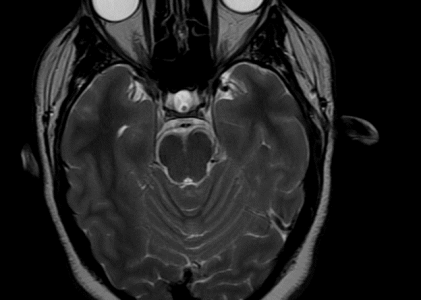
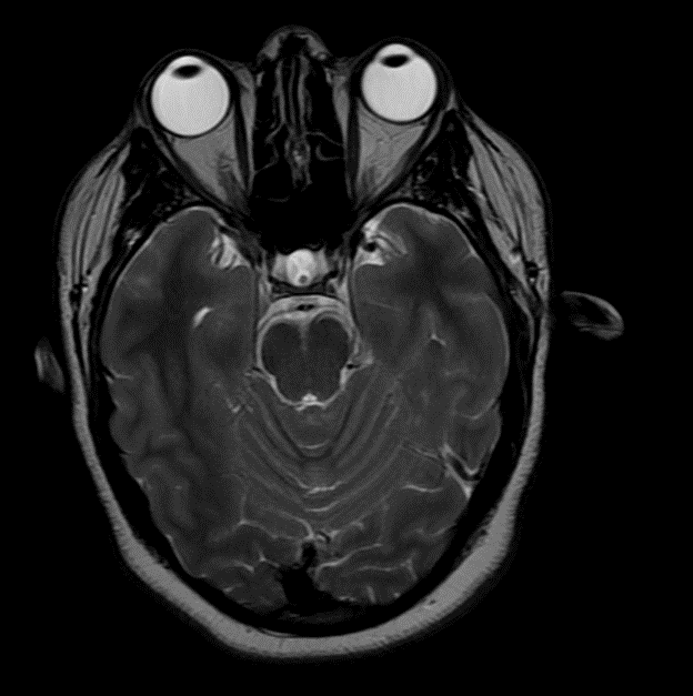
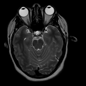
Brain: There is no infarct, hemorrhage, mass or hydrocephalus. There is no enhancing lesion.
Diffusion Images: No evidence of acute ischemic injury.
Pituitary: There is no sellar lesion.
Vasculature: Normal vascular flow voids are demonstrated.
IACS: The internal auditory canals are normal.
Sinuses: The paranasal sinuses appear unremarkable.
There is minor prominence of some lymph nodes in the left parotid gland and in the left suboccipital region.
ORBITS: The globes and extraocular muscles are normal. The optic nerves and optic chiasm are normal. No retro-orbital masses or enhancing lesions are seen. The sella and cavernous sinuses are normal in appearance.
IMPRESSION:
Unremarkable appearance of the brain and orbits.
MR VENOGRAPHY: 6/2/2022 1 :23 PM
INDICATIONS: Papilledema
Report
Technique: Two-dimensional MR venography was performed, followed by 3-dimensional contrast-enhanced time 0 flight MR venography, at 1.5 Tesla . 20 cc Dotarem administered intravenously
COMPARISON: None.
FINDINGS:
No evidence of dural venous sinus thrombosis. There is narrowing of the distal transverse sinuses bilaterally. On the right, the transverse sinus is narrowed by 50%. On the left, the transverse sinus is narrowed by 75%. The dural venous sinus conduit is 2 out of 4 on the right and 1 out of 4 on the left.
IMPRESSION: No dural venous sinus thrombosis. Abnormal combined dural venous conduit score of 3 out of 8 consistent with pseudotumor cerebri or idiopathic intracranial hypertension.