
 HISTORY: STATUS POST PATELLA DISLOCATION – UNABLE TO DORSIFLEX ANKLE
COMPARISON: None.
TECHNIQUE: Multiplanar images of the left knee were obtained at 1.5 Tesla without IV contrast.
LIGAMENTS:
ACL: The anterior cruciate ligament is intact.
PCL: The posterior cruciate ligament is intact.
MCL: The medial collateral ligament is intact.
LCL: The lateral collateral ligament is normal.
MENISCI:
Medial Meniscus: The medial meniscus is intact.
Lateral Meniscus: The lateral meniscus is intact.
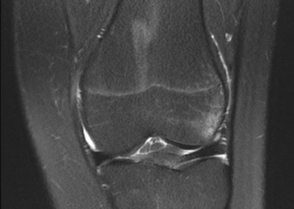
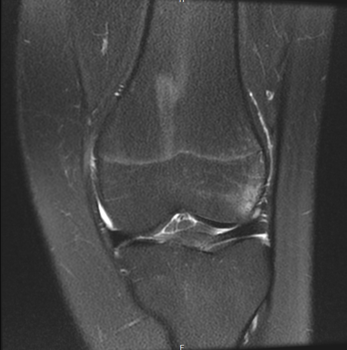
BONES: The patient is recently status post dislocation of the patella, with bone marrow edema in the medial aspect of the patella and within the lateral femoral condyle anteriorly, representing bone contusions related to recent impaction of the patella on the distal femur anteriorly. No significant arthropathy is seen.
SOFT TISSUES: The quadriceps and patellar tendons are intact. There is edema within the medial patellar retinaculum, related to a partial tear, with some edema in the infrapatellar fat pad. There is no significant joint effusion.
IMPRESSION: Status post recent patellar dislocation, with corresponding bone contusions of the patella medially and of the lateral femoral condyle anteriorly. Partial tear of the medial patellar retinaculum. No meniscal or ligamentous tear.