Reason For Exam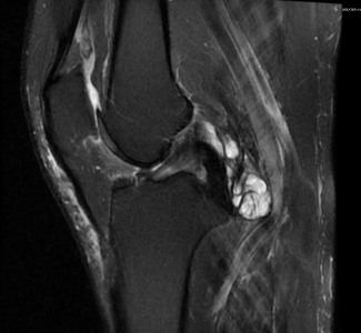
(MR Knee wo Left Visionaire) UNILATERAL PRIMARY OSTEOARTHRITIS OF LEFT KNEE
Report MR LEFT KNEE:
HISTORY: UNILATERAL PRIMARY OSTEOARTHRITIS OF LEFT KNEE
COMPARISON: None
TECHNIQUE: Multiplanar images of the left knee were obtained at 1.5 Tesla without IV contrast. During this public health emergency, we are using enhanced sterilization processes, social distancing measures and PPE for your protection.
LIGAMENTS:
ACL: The anterior cruciate ligament is intact.
PCL: The posterior cruciate ligament is intact.
MCL: The medial collateral ligament is intact.
LCL: The lateral collateral ligament is normal.
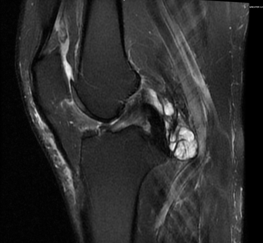
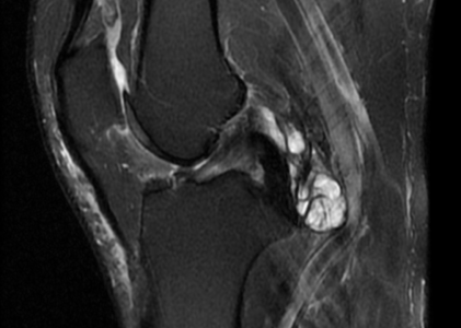
MENISCI: Medial Meniscus: There are broad horizontal tears of the inferior surface of the body and posterior horn of the medial meniscus, extending posteriorly to the meniscal root. Lateral Meniscus: There is a small tear of the inferior surface of the posterior horn of the lateral meniscus.
BONES: There are severe degenerative changes of the medial femorotibial joint, with extensive loss of articular cartilage along the medial femoral condyle and medial tibial plateau. There are prominent medial marginal osteophytes, with smaller osteophytes laterally. There are mild degenerative changes of the patellofemoral joint. No acute bony injury.
SOFT TISSUES: The quadriceps and patellar tendons are intact. There is a moderate knee joint effusion, with loculated synovial fluid in the soft tissues just posterior to the distal PCL.
IMPRESSION: Severe osteoarthritis of the left knee medially, with extensive tears of the body and posterior horn of the medial meniscus. Small tear of the posterior horn of the lateral meniscus.