

HISTORY: INTERNAL DERANGEMENT LEFT KNEE
COMPARISON: None
TECHNIQUE: Multiplanar images of the left knee were obtained at 1.5 Tesla without IV contrast.
LIGAMENTS:
ACL: The anterior cruciate ligament is intact.
PCL: The posterior cruciate ligament is intact.
MCL: The medial collateral ligament is intact.
LCL: The lateral collateral ligament is normal.
MENISCI:
Medial Meniscus: The medial meniscus is intact.
Lateral Meniscus: The lateral meniscus is intact.
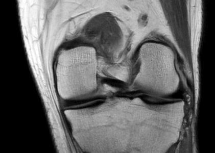
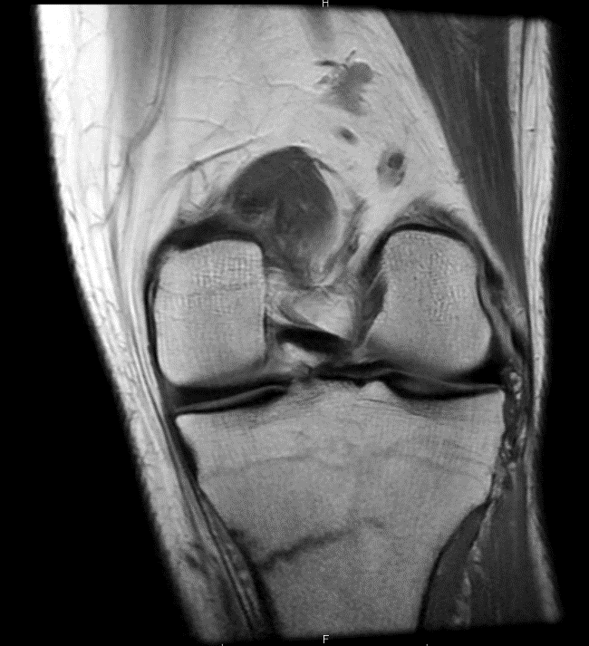
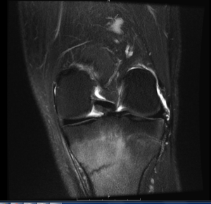
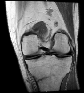
BONES: There is an incomplete transverse fracture involving the medial cortex of the proximal left tibial metaphysis. There is surrounding tibial bone marrow edema. There is mild bone marrow edema within the inferior pole of the patella. The articular surface cartilage along the medial and lateral compartments appears intact. There is fissuring of the articular surface cartilage along the patellar apex.
SOFT TISSUES: There is minimal, focal partial-thickness tear of the proximal patellar tendon. The quadriceps tendon appears normal. The medial and lateral patellar retinacula are intact. There is no significant joint effusion.
IMPRESSION: Stress fracture of the proximal left tibial metaphysis with surrounding bone marrow edema. No ligamentous or meniscal tear.