
Reason For Exam
(MR Knee wo Right) Pain in right knee
Report
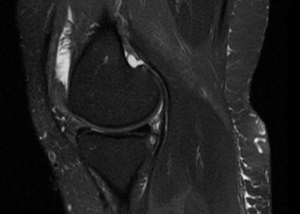
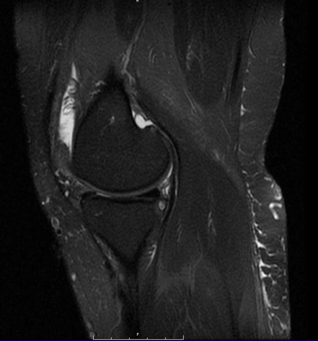
MR RIGHT KNEE:
 HISTORY: Pain in right knee
COMPARISON: None.
TECHNIQUE: Multiplanar images of the right knee were obtained at 1.5 Tesla without IV contrast.
LIGAMENTS:
ACL: The anterior cruciate ligament is intact.
PCL: There is a degenerative posterior cruciate ligament proximally. MCL: The medial collateral ligament is intact.
LCL: The lateral collateral ligament is normal.
MENISCI:
Medial Meniscus: There is a thin horizontal tear of the inferior surface of the posterior horn of the medial meniscus. Lateral Meniscus: The lateral meniscus is intact.
BONES: There are moderate degenerative changes of the medial and lateral femorotibial joints, with more extensive degenerative changes involving the patellofemoral joint, especially along the lateral patellar facet, where there is complete loss of articular cartilage. Subchondral bone marrow edema involves the patella medially and laterally. There is similar bone marrow edema involving the lateral femoral condyle anteriorly.
SOFT TISSUES: The quadriceps and pate!!ar tendons are intact. There is a moderate knee joint effusion.
IMPRESSION: PCL tear proximally. Thin horizontal tear of the inferior surface of the posterior horn of the medial meniscus.
Osteoarthritis of the right knee most significantly involves the patellofemoral joint.