
 HISTORY: INTERVERTERBRAL DISC DEGENERATION AND DISPLACEMENT OF THORACIC REGION – THORACIC RADICULOPATHY
COMPARISON: None
TECHNIQUE: Axial and sagittal images of the thoracic spine were obtained at 1.5 Tesla. During this public health emergency, we are using enhanced sterilization processes, social distancing measures and PPE for your protection
FINDINGS: Alignment is maintained. Vertebral body heights are maintained. No aggressive marrow changes are identified. The thoracic cord is normal in size and signal characteristics. Para-vertebrae soft tissues are unremarkable.
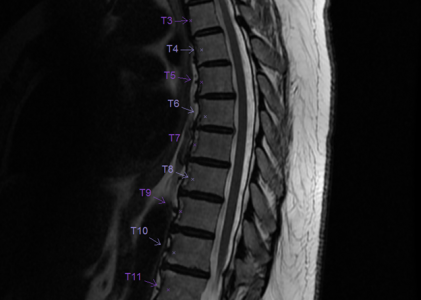
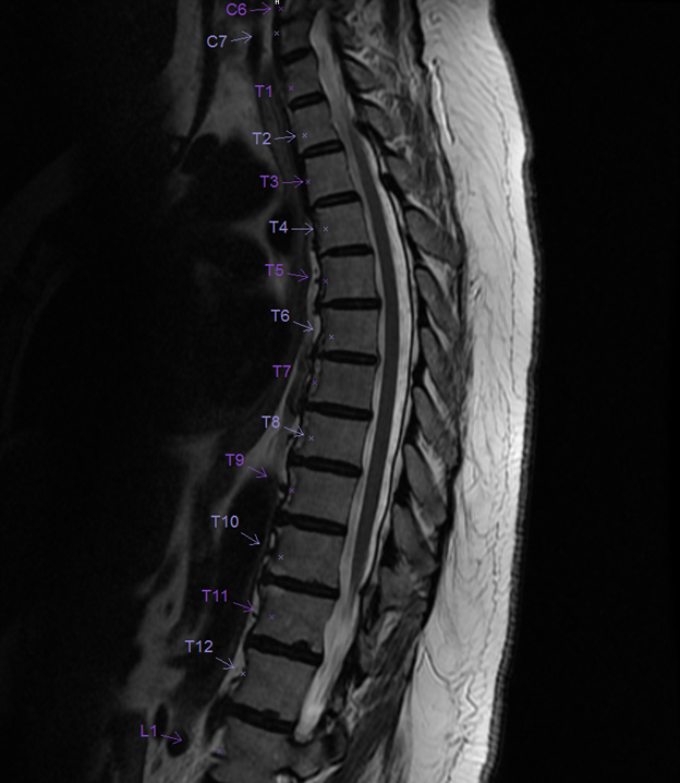
There are minor disc degenerative changes at several levels with small endplate osteophytes. Small central disc protrusions at T5-6 and T6-7 without significant central canal or foraminal stenosis.
IMPRESSION:
Minor thoracic spondylosis with small disc protrusions at C5-6 and C6-7. No significant central canal or foraminal stenosis.