


MRI Knee Case Study
Our case study of the month is an MRI scan of the right knee with history of internal derangement. The MRI Knee Case Study procedure included multi-planar images of the right knee obtained without IV contrast on our 1.5 Tesla MRI machine. There was no comparison scan.
 MRI Exam Findings:

Technique: Multi-planar images of the right knee were obtained at 1.5 Tesla without IV contrast.
Ligaments:
ACL: The anterior cruciate ligament is intact.
PCL: The posterior cruciate ligament is intact.
MCL: The medial collateral ligament is intact.
LCL: The lateral collateral ligament is normal.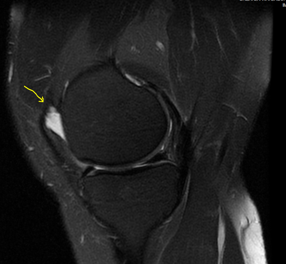
Menisci:
Medial Meniscus: The medial meniscus is intact.
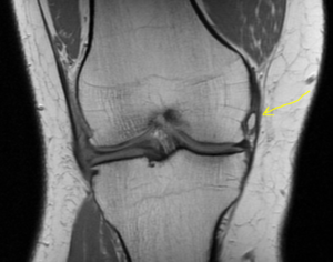
Lateral Meniscus: There is a complex tear along the anterior horn, body and posterior horn of the lateral meniscus.
Bones:
There are mild degernative changes int he lateral compartment, with partial thickness articular surface cartilage thinning and adjacent marginal osteophyte formation. There is no fracture.
Soft Tissues:
The quadriceps and patellar tendons are intact. No soft tissue abnormality is seen. There is no significant joint effusion.
Impression:
Complex lateral meniscal tear.