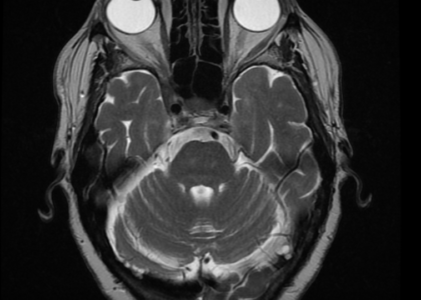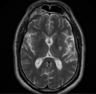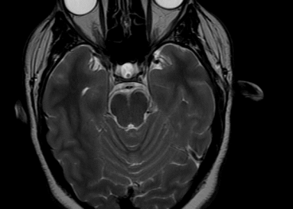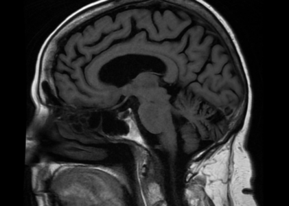
MRI Brain without Contrast
Reason For Exam (MR Brain wo) Other specified injuries of head, initial encounter, Other visua).disturbances. Fall on same level from slipping, tripping and stumbling without subsequent striking against object Report MR HEAD: HISTORY: Other specified injuries of head, initial encounter. Other visual disturbances, Fall on same level from slipping, tripping …