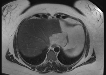
MR Liver Iron Quantification

MR liver iron quantification is a non-invasive means of measuring liver iron concentration, a key indicator in the management of patients with hemochromatosis (primary or secondary). Hemochromatosis is characterized by a progressive increase in total body iron stores, with abnormal iron deposition in multiple organs. Primary hemochromatosis is a genetic …

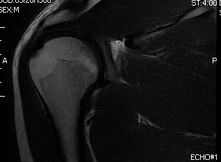
MR LEFT SHOULDER

HISTORY: LEFT SHOULDER ROTATOR CUFF TENDONITIS COMPARISON: None TECHNIQUE: Multiplanar images of the left shoulder were obtained at 1.5 Tesla without IV contrast. FINDINGS: AC JOINT: The acromioclavicular joint appears normal. There is mild narrowing of the subacromial space. There is no significant fluid with in the subacromial/subdeltoid bursa. ROTATOR …

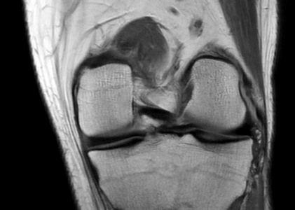
MR LEFT KNEE

HISTORY: INTERNAL DERANGEMENT LEFT KNEE COMPARISON: None TECHNIQUE: Multiplanar images of the left knee were obtained at 1.5 Tesla without IV contrast. LIGAMENTS: ACL:  The anterior cruciate ligament is intact. PCL:  The posterior cruciate ligament is intact. MCL:  The medial collateral ligament is intact. LCL:  The lateral collateral ligament is …

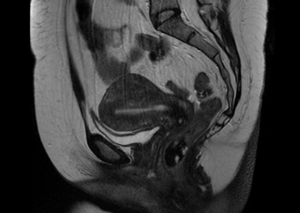
MR PELVIS WITH AND WITHOUT CONTRAST

HISTORY: INTRAMURAL AND SUBSEROUS LEIOMYOMA COMPARISON: CT 11/12/2013 TECHNIQUE: Multiplanar images of the pelvis were obtained at 1.5 Tesla prior to and following administration of 6.5 mL of Gadavist intravenous contrast. FINDINGS: Uterus: The uterus is enlarged measuring 9.3 cm in length. There is a 2.9 cm intramural fibroid of …

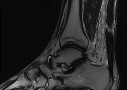
MR RIGHT ANKLE

HISTORY: RIGHT ANKLE PAIN – STATUS POST TRAUMA WITH RIGHT LOWER LEG – ANKLE ECCYMOSIS AND SOFT TISSUE SWELLING COMPARISON: None TECHNIQUE: Multiplanar images of the right ankle were obtained at 1.5 Tesla without IV contrast FINDINGS: BONES: There are degenerative joint changes of the anterior and posterior subtalar joins …