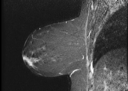
MRI Pelvis Prostate Exam Case Study

MRI Pelvis Prostate Exam with and without IV contrast HISTORY: HIGH GRADE PROSTATIC INTRAEPITHELIAL NEOPLASIA, ELEVATED PROSTATE SPECIFIC ANTIGEN COMPARISON: CT 6/26/2013 TECHNIQUE: Multiplanar images of the pelvis were obtained at 1.5 Tesla prior to and following administration of 8 ml. of Gadavist intravenous contrast. FINDINGS: Urinary Bladder: The urinary …

CASE STUDY HEAD AND ORBITS

MR HEAD AND ORBITS WITH AND WITHOUT CONTRAST HISTORY: Headaches – Blurry Vision COMPARISON: 9/12/2013 TECHNIQUE: Multiplanar images of the head were obtained at 1.5 Tesla prior to and following administration of 6 mL of Gradavist intravenous contrast.  Thin sections through orbits were also obtained. FINDINGS:   HEAD:: There are mild hypertrophic degenerative …

MR RIGHT SHOULDER

CASE STUDY: MR RIGHT SHOULDER HISTORY: SEVERE BILATERAL SHOULDER PAIN COMPARISON: None TECHNIQUE: Multiplanar images of the right shoulder were obtained at 1.5 Tesla without IV contrast FINDINGS: AC JOINT: There are mild hypertrophic degenerative changes of the acromioclavicular joint. There is mild narrowing of the subacromial space. There is …

MRI Thoracic Spine Case Study

MRI Thoracic Spine Case Study HISTORY:  Trauma COMPARISON: CT 5/28/2018 TECHNIQUE:  Axial and sagittal images of the thoracic spine were obtained at 1.5 Tesla FINDINGS: As noted on the recent CT scan of the chest, there is a compression fracture of the T8 vertebral body, with loss of vertebral height …

MRI LEFT ELBOW

Our Case Study of the Month is an: MR LEFT ELBOW HISTORY: LEFT ELBOW PAIN SINCE AUGUST 2017 AFTER STRAINING ARM COMPARISON: None TECHNIQUE: Multiplanar images of the left elbow were obtained at 1.5 Tesla without IV contrast FINDINGS: BONES: There is no fracture or subluxation. No areas of bone marrow …

MR Abdomen With and Without Contrast

HISTORY: RIGHT LOBE MASS, HISTORY OF CIRRHOSIS COMPARISON: Abdominal ultrasound 6/22/12 TECHNIQUE: Multiplanar images of the abdomen were obtained at 1.5 Tesla prior to and following administration of IV contrast. FINDINGS: Liver: The liver is diffusely heterogeneous nodular in contour consistent with cirrhosis. There are no enhancing liver masses seen. …

MRI of the Pelvis Case Study

MRI of the Pelvis COMPARISON: MRI of Pelvis 11/06/2017 TECHNIQUE: Multiplanar images of the pelvis were obtained at 1.5 Tesla prior to and following administration of 8 mL of Gadavist intravenous contrast. FINDINGS: GI TRACT: There is a concentric, heterogeneous lobular mass along the wall of the rectosigmoid junction extending …

MRI of the Chest

MRI Of the Chest HISTORY:  Soft Tissue Mass – Leff Trapezius Muscle COMPARISON:  None TECHNIQUE:  Multiplanar images of the chest were obatined at 1.5 Tesla prior to and following administration of IV contrast. CONTRAST:  8 mL of Gadavist Intravenous contrast. FINDINGS: There is a fat-containing nonenhancing lesion along the posterior left trapezius muscle medially …